Lateral rectus muscle
| Lateral rectus | |
|---|---|
 Lateral rectus muscle: is shown in this superior view of the eye. The lateral rectus is on the right side of the image. | |
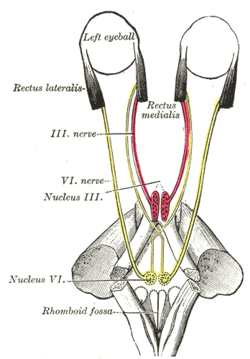 Figure showing the mode of innervation of the Recti medialis and lateralis of the eye. | |
| Details | |
| Latin | musculus rectus lateralis bulbi |
| annulus of Zinn at the orbital apex | |
| 7 mm temporal to the limbus | |
| abducens nerve | |
| Actions | abducts the eyeball (makes it move outwards) |
| Identifiers | |
| Gray's | p.1022 |
| Dorlands /Elsevier | m_22/12550497 |
| TA | A15.2.07.013 |
| FMA | 49038 |
| Anatomical terms of muscle | |
- For the muscle of the neck, see Rectus capitis lateralis muscle
The lateral rectus muscle is a muscle in the orbit. It is one of six extraocular muscles that control the movements of the eye (abduction in this case). Its function is to bring the pupil away from the midline of the body.
Structure
The lateral rectus originates at the lateral part of the annulus of Zinn, also known as the annular tendon or common tendinous ring, and inserts into the temporal side of the eyeball. The annulus of Zinn is a tendinous ring that surrounds the optic nerve and serves as the origin for four of the six extraocular muscles, excluding the inferior oblique muscle and superior oblique muscle.[1]
Innervation
It is the only muscle innervated by the abducens nerve, cranial nerve VI.
Function
Clinical significance
A sixth nerve palsy, also known as abducens nerve palsy, is a neurological defect that results from a damaged or impaired abducens nerve. This defect can result in horizontal double vision and reduced lateral movement.[2] Proper function of the lateral rectus is tested clinically by asking the patient to look laterally.
Additional images
-

Lateral rectus muscle
-

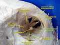
Dissection showing origins of right ocular muscles, and nerves entering by the superior orbital fissure.
-

Lateral view of the eyeball with lateral rectus muscle visible (cut).
-
Lateral rectus muscle
-
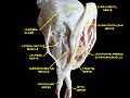
Extrinsic eye muscle. Nerves of orbita. Deep dissection.
-

Extrinsic eye muscle. Nerves of orbita. Deep dissection.
-

Extrinsic eye muscle. Nerves of orbita. Deep dissection.
-

Extrinsic eye muscle. Nerves of orbita. Deep dissection.
External links
- -66715569 at GPnotebook
- Anatomy figure: 29:01-05 at Human Anatomy Online, SUNY Downstate Medical Center
- Cranial Nerves at Yale 6-1
- lateral+rectus+muscle at eMedicine Dictionary
| ||||||||||||||||||||||||||||||||||||||||||||||||||||||||||||||||||||||||||