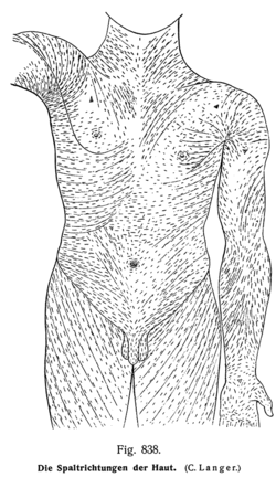
Langer's lines
| Langer's lines | |
|---|---|
 | |
| Identifiers | |
| TA | A16.0.00.008 |
| FMA | 71951 |
| Anatomical terminology | |
Langer's lines, sometimes called cleavage lines, are topological lines drawn on a map of the human body. They were historically defined by the direction in which the skin of a human cadaver will split when struck with a spike. They correspond to the natural orientation of collagen fibers in the dermis, and are generally parallel to the orientation of the underlying muscle fibers. Langer's lines have relevance to forensic science and the development of surgical techniques.
History
The lines were first discovered in 1861 by Austrian anatomist Karl Langer (1819-1887),[1][2][3] though he cited the surgeon Baron Dupuytren as being the first to recognise the phenomenon. Langer punctured numerous holes at short distances from each other into the skin of a cadaver with a tool that had a circular-shaped tip, similar to an ice pick. He noticed that the resultant punctures in the skin had ellipsoidal shapes. From this testing he observed patterns and was able to determine "line directions" by the longer axes of the ellipsoidal holes and lines .
Application
Knowing the direction of Langer's lines within a specific area of the skin is important for surgical operations, particularly cosmetic surgery. If a surgeon has a choice about where and in what direction to place an incision, he or she may choose to cut in the direction of Langer's lines. Incisions made parallel to Langer's lines may heal better and produce less scarring than those that cut across. Conversely, incisions perpendicular to Langer's lines have a tendency to pucker and remain obvious, although sometimes this is unavoidable. Keloids are more common when incision is given across Langer's lines. Sometimes the exact direction of the collagen fibers are unknown, because in some regions of the body there are differences between different individuals. Also, the lines described by Kraissl differ in some ways from Langer's lines, particularly on the face.
The orientation of stab wounds relative to Langer's lines can have a considerable impact upon the presentation of the wound.[4]
Alternatives
Other authors have created topological skin maps. Kraissl's lines differ from Langer's lines in that while Langer's lines were defined in cadavers,[5] Kraissl's lines have been defined in living individuals. Also, the method used to identify Kraissl's lines is not traumatic.
Langers line perpendicular to muscle fibrea not parallel References== grabb smith latest edition
- ↑ Karl Langer, "Zur Anatomie und Physiologie der Haut. Über die Spaltbarkeit der Cutis". Sitzungsbericht der Mathematisch-naturwissenschaftlichen Classe der Wiener Kaiserlichen Academie der Wissenschaften Abt. 44 (1861)
- ↑ Langer, K (January 1978). "On the anatomy and physiology of the skin". British Journal of Plastic Surgery 31 (1): 3–8. doi:10.1016/0007-1226(78)90003-6. PMID 342028.
- ↑ "Method and apparatus for determining the lines of optimal direction for surgical cuts in the human skin - US Patent 6418339".
- ↑ "Forensic Pathology".
- ↑ Wilhelmi BJ, Blackwell SJ, Phillips LG (July 1999). "Langer's lines: to use or not to use". Plast. Reconstr. Surg. 104 (1): 208–14. doi:10.1097/00006534-199907000-00032. PMID 10597698.