Lamina affixa
| Lamina affixa | |
|---|---|
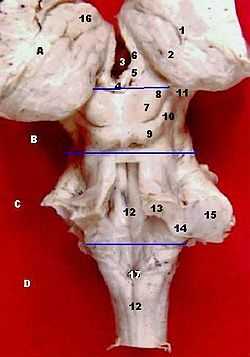 1. Taenia choroidea (and lateral: Lamina affixa, Stria terminalis) 2. Thalamus, Pulvinar thalami 3. Third ventricle 4. Stalk of pineal gland 5. Habenula 6. Stria medullaris 7. Superior colliculus 8. Brachium of superior colliculus 9. Inferior colliculus 10. Brachium of inferior colliculus 11. Medial geniculate nucleus 12. Sulcus medianus 13. Superior cerebellar peduncles 14. Inferior cerebellar peduncle 15. Middle cerebellar peduncles 16. Tuberculum anterius thalami 17. Obex, Area postrema | |
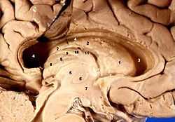 Human brain left dissected midsagittal view (Lamina affixa is #10) | |
| Details | |
| Latin | lamina affixa |
| Identifiers | |
| Gray's | p.838 |
| Dorlands /Elsevier | l_02/12475860 |
| TA | A14.1.09.276 |
| FMA | 83709 |
| Anatomical terms of neuroanatomy | |
Lamina affixa is a layer of epithelium growing on the surface of the thalamus and forming the floor of the central part of lateral ventricle, on whose medial margin is attached the choroid plexus of the lateral ventricle; it covers the thalamostriate and choroidal veins. The torn edge of this plexus is called the choroid tenia.
On the surface of the terminal vein is a narrow white band, named the lamina affixa.
GDF-15/MIC-1 has been observed in lamina affixa cells.[1]
References
This article incorporates text in the public domain from the 20th edition of Gray's Anatomy (1918)
- ↑ Schober A, Böttner M, Strelau J et al. (October 2001). "Expression of growth differentiation factor-15/ macrophage inhibitory cytokine-1 (GDF-15/MIC-1) in the perinatal, adult, and injured rat brain". J. Comp. Neurol. 439 (1): 32–45. doi:10.1002/cne.1333. PMID 11579380.
External links
| ||||||||||||||||||||||||||||||||||||||||||||||||||||||||||||||||||||||||||||
| ||||||||||||||||||||||||||||