Internal ribosome entry site
An internal ribosome entry site, abbreviated IRES, is a nucleotide sequence that allows for translation initiation in the middle of a messenger RNA (mRNA) sequence as part of the greater process of protein synthesis. Usually, in eukaryotes, translation can be initiated only at the 5' end of the mRNA molecule, since 5' cap recognition is required for the assembly of the initiation complex. The location for these sites is often in the 5'UTR, but can occur in many different places in an mRNA.
History
These sequences were first discovered in 1988 in poliovirus RNA and encephalomyocarditis virus RNA in the labs of Nahum Sonenberg[1] and Eckard Wimmer,[2] respectively. They are described as distinct regions of RNA molecules that are able to attract the eukaryotic ribosome to the mRNA molecule and, therefore, allow translation initiation to occur. This process became known as the internal initiation of translation. It has been hypothesized that IRES elements have a distinct secondary or even tertiary structure, but similar structural features at the levels of either primary or secondary structure that are common to all IRES segments have not been reported to date.
In recent years it has become common for molecular biologists to insert IRES sequences into their vectors to allow for expression of two genes from a single vector-- for example, a transgene and a fluorescent reporter molecule. The first gene is initiated at the normal 5' cap, and the second at the IRES.
Location
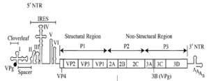
It is common that IRESes are located in the 5'UTR of RNA viruses and allow translation of the RNAs in a cap-independent manner. However, mRNAs of viruses from Dicistroviridae family possess two ORFs (open reading frames), translation of both being directed by two distinct IRESes. It was later suggested that some mammalian mRNAs also have IRES. Several cellular IRES elements are thought to be located in eukaryotic mRNAs encoding genes involved in stress survival, and other processes critical to survival. As of September 2009, there are 60 animal and 8 plant viruses reported to contain IRES segments and 115 mRNA sequences containing them as well.[3]
Activation
IRES are often used by viruses as a means to ensure that viral translation is active during periods of time when host translation is inhibited. These mechanisms of host translation inhibition are varied, and can be initiated by both virus and host, depending on the type of virus in question. However, in the case of most picornaviruses, this is accomplished by the viral protease cleaving eIF-4G so that it cannot interact with eIF-4E. Interaction between these two initiation factors is necessary for mRNA 5'cap to 3'poly-A-tail loop formation, which is usually a necessary event for initiation of translation. The virus may even use the eIF-4G to aid in initiation of IRES-mediated translation.
The cell may also use IRES to increase translation of certain proteins during mitosis and programmed cell death. In mitosis, the cell dephosphorylates eIF-4E so that it has little affinity for the 5'cap. As a result, the pre-initiation mRNA loop is not formed, and the translational machinery is diverted to IRES within the mRNA. Many proteins involved in mitosis are encoded by IRES mRNA. In programmed cell death, cleavage of eIF-4G, such as performed by viruses, decreases translation. Lack of essential proteins contributes to the death of the cell, as does translation of IRES mRNA sequences coding proteins involved in controlling cell death.[4]
Mechanism
The mechanism of viral IRES function to date is better characterized than the mechanism of cellular IRES function,[5] for which no clear mechanism have been proposed yet. Hepatitis C Virus-related IRESs directly bind 40S ribosomal subunit in such a way that their initiator codons are located in ribosomal P-site without mRNA scanning. These IRESs do not require Eukaryotic initiation factors eIF1, 1A, 4A, 4B, and 4E. Picornavirus IRES do not attract 40S directly, but rather through high-affinity eIF4G-binding site.[6] In addition, many viral IRES (as well as cellular IRES) require additional proteins to mediate their function, known as IRES trans-acting factors (ITAFs). The role of ITAFs in IRES function is currently the subject of intense research.
Testing
Testing a particular RNA sequence for IRES activity relies on a bicistronic reporter construct. When an IRES segment is located between two reporter open reading frames in a eukaryotic mRNA molecule (a bicistronic mRNA), it can drive translation of the downstream protein coding region independently of the 5'-cap structure bound to the 5' end of the mRNA molecule. In such a setup both proteins are produced in the cell. The first reporter protein located in the first cistron is synthesized by the cap-dependent initiation approach while translation initiation of the second protein is directed by the IRES segment located in the intercistronic spacer region between the two reporter protein coding regions. However, there are several caveats to be aware of when interpreting data produced using bicistronic reporter constructs.[7] For example, there are several known cases of mis-reported IRES segments that were later recognized as promoter-containing regions. More recently, splice acceptor sites within several presumed IRES segments have been shown to be responsible for apparent IRES function in bicistronic reporter assays.[8]
Types
| Virus | IRES |
| Poliovirus | Picornavirus IRES |
| Rhinovirus | Picornavirus IRES |
| Encephalomyocarditis virus | Picornavirus IRES |
| Foot-and-mouth disease virus | Aphthovirus IRES |
| Hepatitis A virus | Hepatitis A IRES |
| Hepatitis C virus | Hepatitis C IRES |
| Classical swine fever virus | Pestivirus IRES |
| Bovine viral diarrhea virus | Pestivirus IRES |
| Friend murine leukemia | |
| Moloney murine leukemia (MMLV) | |
| Rous sarcoma virus | |
| Human immunodeficiency virus | |
| Plautia stali intestine virus | Cripavirus internal ribosome entry site (IRES) |
| Rhopalosiphum padi virus | Cripavirus internal ribosome entry site (IRES) |
| Cricket paralysis virus | Cripavirus internal ribosome entry site (IRES) |
| Triatoma virus | Cripavirus internal ribosome entry site (IRES) |
| Kaposi's sarcoma-associated herpesvirus | Kaposi's sarcoma-associated herpesvirus IRES |
| Marek's disease virus MDV | 5'Leader IRES and intercistronic IRES in the 1.8-kb family of immediate early transcripts (IRES)1 |
| Protein type | Proteins |
| Growth factors | Fibroblast growth factor (FGF-1 IRES and FGF-2 IRES), Platelet-derived growth factor B (PDGF/c-sis IRES), Vascular endothelial growth factor (VEGF IRES), Insulin-like growth factor 2 (IGF-II IRES) |
| Transcription factors | Antennapedia, Ultrabithorax, MYT-2, NF-κB repressing factor NRF, AML1/RUNX1, Gtx homeodomain protein |
| Translation factors | Eukaryotic initiation factor 4G (elF4G)a, Eukaryotic initiation factor 4Gl (elF4Gl)a, Eukaryotic translation initiation factor 4 gamma 2 (EIF4G2,DAP5) |
| Oncogenes | c-myc, L-myc, Pim-1, Protein kinase p58PITSLRE, p53 |
| Transporters/receptors | Cationic amino acid transporter (SLC7A1,Cat-1), Nuclear form of Notch 2, Voltage-gated potassium channel |
| Activators of apoptosis | Apoptotic protease activating factor (Apaf-1) |
| Inhibitors of apoptosis | X-linked inhibitor of apoptosis (XIAP), HIAP2, Bcl-xL, Bcl-2 |
| Proteins localized in neuronal dendrites | Activity-regulated cytoskeletal protein (ARC), α-subunit of calcium calmodulin dependent kinase II dendrin, Microtubule-associated protein 2 (MAP2), neurogranin (RC3), Amyloid precursor protein |
| Others | Immunoglobulin heavy chain binding protein (BiP), Heat shock protein 70, β-subunit of mitochondrial H+-ATP synthase, Ornithine decarboxylase, connexins 32 and 43, HIF-1α, APC |
References
- ↑ Pelletier J, Sonenberg N (1988). "Internal initiation of translation of eukaryotic mRNA directed by a sequence derived from poliovirus RNA". Nature 334 (6180): 320–5. doi:10.1038/334320a0. PMID 2839775.
- ↑ Jang SK, Kräusslich HG, Nicklin MJ, Duke GM, Palmenberg AC, Wimmer E (August 1988). "A segment of the 5' nontranslated region of encephalomyocarditis virus RNA directs internal entry of ribosomes during in vitro translation". J. Virol. 62 (8): 2636–43. PMC 253694. PMID 2839690.
- ↑ Mokrejs M, Vopálenský V, Kolenaty O et al. (January 2006). "IRESite: the database of experimentally verified IRES structures (www.iresite.org)". Nucleic Acids Res. 34 (Database issue): D125–30. doi:10.1093/nar/gkj081. PMC 1347444. PMID 16381829.
- ↑ Alberts, B., Johnson, A., Lewis, J., Raff, M., Roberts, K., Walter, P. (2002). Molecular Biology of the Cell. Garland Science. pp. 447–448. ISBN 0-8153-4072-9.
- ↑ López-Lastra M, Rivas A, Barría MI (2005). "Protein synthesis in eukaryotes: the growing biological relevance of cap-independent translation initiation". Biol. Res. 38 (2–3): 121–46. doi:10.4067/s0716-97602005000200003. PMID 16238092.
- ↑ 6.0 6.1 6.2 Hellen CU, Sarnow P (2001). "Internal ribosome entry sites in eukaryotic mRNA molecules". Genes Dev. 15 (13): 1593–612. doi:10.1101/gad.891101. PMID 11445534.
- ↑ Kozak M (2005). "A second look at cellular mRNA sequences said to function as internal ribosome entry sites". Nucleic Acids Res. 33 (20): 6593–602. doi:10.1093/nar/gki958. PMC 1298923. PMID 16314320.
- ↑ Baranick BT, Lemp NA, Nagashima J, Hiraoka K, Kasahara N, Logg CR (March 2008). "Splicing mediates the activity of four putative cellular internal ribosome entry sites". Proc. Nat. Acad. Sci. 105 (12): 4733–38. doi:10.1073/pnas.0710650105. PMC 2290820. PMID 18326627.
External links
- IRESite
- Malys N, McCarthy JEG (2010). "Translation initiation: variations in the mechanism can be anticipated". Cellular and Molecular Life Sciences 68 (6): 991–1003. doi:10.1007/s00018-010-0588-z. PMID 21076851.