Internal arcuate fibers
| Internal arcuate fibers | |
|---|---|
| Identifiers | |
| NeuroLex ID | Internal arcuate fiber bundle |
| TA | A14.1.04.109 |
| FMA | 72629 |
| Anatomical terms of neuroanatomy | |
| Internal arcuate fibers | |
|---|---|
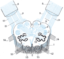
 Diagram showing the course of the arcuate fibers. (Testut.) 1. Medulla oblongata anterior surface. 2. Anterior median fissure. 3. Fourth ventricle. 4. Inferior olivary nucleus, with the accessory olivary nuclei. 5. Gracile nucleus. 6. Cuneate nucleus. 7. Trigeminal. 8. Inferior peduncles, seen from in front. 9. Posterior external arcuate fibers. 10. Anterior external arcuate fibers. 11. Internal arcuate fibers(no it's actually olivo-cerebellar tracts). 12. Peduncle of inferior olivary nucleus. 13. Nucleus arcuatus. 14. Vagus. 15. Hypoglossal. | |
 Section of the medulla oblongata at about the middle of the olive. (Arcuate fibers labeled at center right.) | |
| Details | |
| Latin | fibrae arcuatae internae |
| Identifiers | |
| Gray's | p.782 |
| Dorlands /Elsevier | f_05/12361842 |
| TA | A14.1.04.109 |
| FMA | 72629 |
| Anatomical terminology | |
Internal arcuate fibers are the axons of second-order neurons contained within the gracile and cuneate nuclei of the medulla oblongata.
These fibers cross (decussate) from one side of the medulla to the other to form the medial lemniscus.
Part of the posterior column-medial lemniscus system (second neuron), the internal arcuate fibers are important for relaying the sensation of fine touch and proprioception to the thalamus and ultimately to the cerebral cortex.
External links
- Hier-792 at NeuroNames
- Photo at Indiana.edu
| ||||||||||||||||||||||||||||||||||||||||||||||||||||||||||||||||