Inferior orbital fissure
| Inferior orbital fissure | |
|---|---|
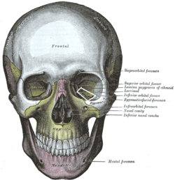 The skull from the front. (Label for inferior orbital fissure is at center right.) | |
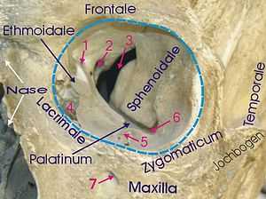 1 Foramen ethmoidale, 2 Canalis opticus, 3 Fissura orbitalis superior, 4 Fossa sacci lacrimalis, 5 Sulcus infraorbitalis, 6 Fissura orbitalis inferior, 7 Foramen infraorbitale | |
| Details | |
| Latin | Fissura orbitalis inferior |
| Identifiers | |
| Gray's | p.189 |
| Dorlands /Elsevier | f_08/12365602 |
| TA | A02.1.00.084 |
| FMA | 54802 |
| Anatomical terminology | |
The lateral wall and the floor of the orbit are separated posteriorly by the inferior orbital fissure which transmits the zygomatic branch of the maxillary nerve, and the ascending branches from the pterygopalatine ganglion. The infraorbital vessels are found in the inferior orbital fissure, and travel down the infraorbital groove into the infraorbital canal and exit through the infraorbital foramen.
It is formed by the sphenoid bone and maxilla.
Images
-
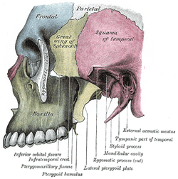
Left infratemporal fossa.
-

Horizontal section of nasal and orbital cavities.
-

Dissection showing origins of right ocular muscles, and nerves entering by the superior orbital fissure.
-
Inferior orbital fissure.
See also
- Foramina of skull
References
This article incorporates text in the public domain from the 20th edition of Gray's Anatomy (1918)
External links
| Wikimedia Commons has media related to Inferior orbital fissure. |
- lesson3 at The Anatomy Lesson by Wesley Norman (Georgetown University) (orbitforamina) (#3)
- Anatomy diagram: 34256.000-1 at Roche Lexicon - illustrated navigator, Elsevier
- Anatomy diagram: 34257.000-1 at Roche Lexicon - illustrated navigator, Elsevier
| ||||||||||||||||||||||||||||||||||||||||||||||||||||||||||||||||||||||||||||||||||||||||||||||||||||||