Impalefection

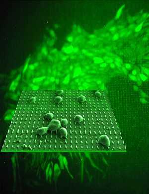
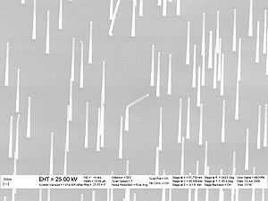
Impalefection is a method of gene delivery using nanomaterials, such as carbon nanofibers, carbon nanotubes, nanowires.[1] Needle-like nanostructures are synthesized perpendicular to the surface of a substrate. Plasmid DNA containing the gene, intended for intracellular delivery, is attached to the nanostructure surface. A chip with arrays of these needles is then pressed against cells or tissue. Cells that are impaled by nanostructures can express the delivered gene(s).
As one of the types of transfection, the term is derived from two words - impalement and infection.
Applications
One of the features of impalefection is spatially resolved gene delivery that holds potential for such tissue engineering approaches in wound healing as gene activated matrix technology.[2]
Carrier materials
Vertically aligned carbon nanofiber arrays prepared by photolithography and plasma enhanced chemical vapor deposition are one of the suitable types of material. Silicon nanowires is another choice of nanoneedles that have been utilized for impalefection.

See also
References
- ↑ McKnight, T.E., A.V. Melechko, D.K. Hensley, D.G.J. Mann, G.D. Griffin, and M.L. Simpson (2004). "Tracking gene expression after DNA delivery using spatially indexed nanofiber arrays". Nano Letters 4 (7): 1213–1219. doi:10.1021/nl049504b.
- ↑ Bonadio J. (2000). "Local gene delivery for tissue regeneration". J Regener Med 1: 25–29. doi:10.1089/152489000414552.