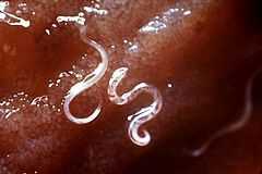
Helminths

Helminths (/ˈhɛlmɪnθs/), also commonly known as parasitic worms, are large multicellular organisms, which when mature can generally be seen with the naked eye. They are often referred to as intestinal worms even though not all helminths reside in the intestines; for example Schistosomes are not intestinal worms, but rather reside in blood vessels.
There is no clear consensus on the taxonomy of helminths, it is more of a commonly used term to describe certain worms with superficial similarities. These are plathelminths (cestodes and trematodes) and nemathelminths (nematodes) - both of these are parasitic worm types – and the annelida, which is not parasitic or at the most ectoparasites like the leeches.[1]
Many, but not all, of the worms referred to as helminths belong to the group of intestinal parasites. An infection by a helminth is known as helminthiasis, soil-transmitted helminthiasis, helminth infection or intestinal worm infection. The same naming convention applies to all helminths whereby the ending "-asis" (or in veterinary science the ending "-osis") at the end of the name of the worm is added to signify the infection with that particular worm, e.g. Ascaris is the name of a particular helminth, and Ascariasis is the name of the infectious disease caused by this helminth.
Helminths are worm-like organisms living in and feeding on living hosts, receiving nourishment and protection while disrupting their hosts' nutrient absorption, causing weakness and disease. Those that live inside the digestive tract are called intestinal parasites. They can live inside humans and other animals. In their adult form, helminths cannot multiply in humans.[2] Helminths are able to survive in their mammalian hosts for many years due to their ability to manipulate the immune response by secreting immunomodulatory products.[3] Helminth ova (or eggs) have a strong shell that protects the eggs against a range of environmental conditions.
Helminthology is the study of parasitic worms and their effects on their hosts. The word helminth comes from Greek hélmins, a kind of worm.
Taxonomy


There is no real consensus on the taxonomy (or groupings) of the helminths, particularly with the nematodes.[4] The term "helminth" contains a number of phyla, many of which are completely unrelated. However, for practical considerations the term is still used nowadays to describe four groups with superficial similarities, the phyla Annelida, Platyhelminths, Nematoda and Acanthocephala.[4]
There is in fact no helminth classification, it is an “artificial” term.[5][6]
The most important helminths in the sanitation field are the human parasites, which is why most people relate the term helminth to them, where they are classified as nemathelminthes (nematodes) and platyhelminthes, depending on whether they possess a round or flat-shaped body respectively. The latter are further divided into cestodes and trematodes depending on whether or not they have a segmented body.[7]
Ringworm (dermatophytosis) is actually caused by various fungi and not by a parasitic worm.
Common characteristics
Helminths are a group of evolutionary unrelated organisms which share a similar form. Helminths include members of the following taxa: monogeneans, cestodes (tapeworms), nematodes (roundworms), and trematodes (flukes). The number of species of the different helminth types is vast, in the order of 1 million. The nematodes are the most diverse of all the helminths with the highest number of species.
Characteristics that are common for all helminths include:
Life time:
- The life time of adult worms varies tremendously from one species to another but is generally in the range of 1 to 8 years (see following table). This life time of several years is a result of their ability to manipulate the immune response of their hosts by secreting immunomodulatory products.[3]
- Helminths can be either hermaphrodites (i.e. can have both sexes), like tapeworms and the flukes (except the blood fluke which is not a hermaphrodite), or have their sexes differentiated, like the roundworms.
Eggs:
- All helminths produce eggs (also called ova) for reproduction.
- Helminth eggs have a strong shell that protects them against a range of environmental conditions. This shell consists of three layers: a lipoidal inner layer, a chitinous middle layer and outer proteinic layer.[8]
- Generally thousands or even hundreds of thousands of eggs are produced each time the female worm deposits its eggs - a process called ovoposition. The following table shows a large variation in the amount of eggs produced by the different worms in one event; it varies in the range of 3,000 to 700,000.
- The frequency of egg deposition from an adult helminth is generally daily, or up to six times per day for some Taenia species.
- Adult trematodes lay smaller numbers of eggs compared to cestodes or nematodes. However, the egg develops into a miracidia from which thousands of cercariae, swimming larvaes, develop. This means that one egg can produce thousands of adult worms.[9]
- Helminth eggs remain viable for 1-2 months in crops and for many months in soil, fresh water, and sewage, even for several years in faeces, night soil and sludge, a period that is much longer compared to other kind of microorganisms.[10][11]
Larvae:
- Larvae hatch from these eggs (if the eggs are viable), inside or outside the host, depending on the type of helminth. Life cycles of the helminths differ in this and other specific aspects. Eggs that are no longer viable do not produce any larvae.
- The larvae maturing in the host takes from about two weeks up to four months depending on the helminth species.
The following table shows the principal morphological and reproductive distinctions for three helminth groups:
| Tapeworms (Cestodes) | Flukes (Trematodes) | Roundworms (Nematodes) | ||||||
|---|---|---|---|---|---|---|---|---|
| Species (examples) | Taenia solium, Taenia saginata, Hymenolepis spp., Echinoccocus granulosus, Multiceps multiceps | Schistosoma mansoni, Schistosoma japonicum, | Ascaris, Onchocera, Rhabditis, Trichuris, Necator americanus, Anchylostoma duodenale | |||||
| Example diseases in humans | Tapeworm infection | Schistosomiasis, swimmer's itch | Ascariasis, dracunculiasis (guinea worm), elephantiasis, enterobiasis (pinworm), filariasis, hookworm infection (includes Necatoriasis and Ancylostoma duodenale infection), onchocerciasis, trichinosis, trichuriasis (whipworm) | |||||
| Shape | Segmented plane | Unsegmented plane | Cylindrical | |||||
| Body cavity | No | no | Present | |||||
| Body covering | Tegument | Tegument | Cuticle | |||||
| Digestive tube | No | Ends in cecum | Ends in anus | |||||
| Sex | Hermaphroditic | Hermaphroditic, except schistosomes which are dioecious | Dioecious | |||||
| Attachment organs | Sucker or bothridia, and rostellum with hooks | Oral sucker and ventral sucker or acetabulum | Lips, teeth, filariform extremities, and dentary plates | |||||
|
Number of species |
6000[12] | Estimated > 15,000[13] and 9,000[14] registered | 800,000 to 1,000,000 estimated
25,000 registered[13] | |||||
| Number of species known to infect humans | 40[12] | 16[13] | > 12,000[13] | |||||
| Species |
Hookworm |
Toxocara | ||||||
| Lifetime | Larvae formation |
some days (eggs can survive for months)[15] |
9-15 days[12] |
18 days to several weeks[16] |
1-2 days[17] |
15-30 days[18] |
||
| Larvae growth |
After hatching the larvae move to develop into cysticercoid, which can survive for years in an animal[15] |
5-7 weeks as cercariae in snails and longer periods in wet environments as encysted metacercariae[9] |
10-14 days[16] |
5-10 days (after maturing can survive for weeks outside the host)[17] |
60-70 days (from hatching to mature state)[18] |
5-6 days[12] | ||
| Larvae maturing (in host) |
2 months (form cysticercoid to adult)[15] |
3-4 months[9] |
2-3 months[16] |
2-8 weeks[12] (can become dormant for months) |
||||
| Adult worm |
4-6 weeks |
Several years[15] |
8-10 years[12] |
1-2 years[16] |
Several years[17] |
1 year[18] |
||
| Eggs laid per day | 250,000[2] to 700,000[12] | 3,000 to 25,000[13] | 3,000[2] to 250,000[12] | |||||
| Egg deposition | Frequency |
up to 6 times a day[15] |
daily[16] |
daily[17] |
daily[18] |
|||
| Number of eggs per event |
50,000-100,000[15] |
5,000-10,000[12] |
3,000-20,000[18] |
|||||
| Larvae per egg | 1 | 1 | 300 cercariae (Schistosoma), 250,000 metacercariae (Fasciola)[13] | 1 | 1 | 1 | 1 | |
Draft genomes for all categories of helminth have been sequenced in recent years and are available through the ParaSite sub-portal of WormBase.[20]
Use in medicine
Parasitic worms have been used as a medical treatment for various diseases, particularly those involving an overactive immune response.[21] As humans have evolved with parasitic worms, proponents argue they are needed for a healthy immune system.[21] Scientists are looking for a connection between the prevention and control of parasitic worms and the increase in allergies such as hay-fever in developed countries.[21] Parasitic worms may be able to damp down the immune system of their host, making it easier for them to live in the intestine without coming under attack.[21] This may be one mechanism for their proposed medicinal effect.
One study suggests a link between the rising rates of metabolic syndrome in the developed worlds and the largely successful efforts of Westerners to eliminate intestinal parasites. The work suggests eosinophils (a type of white blood cell) in fat tissue play an important role in preventing insulin resistance by secreting interleukin 4, which in turn switches macrophages into "alternative activation". Alternatively-activated macrophages are important to maintaining glucose homeostasis (i.e., blood sugar regulation). Helminth infection causes an increase in eosinophils. In the study, the authors fed rodents a high-fat diet to induce metabolic syndrome, and then injected them with helminths. Helminth infestation improved the rodents' metabolism.[22] The authors concluded:
Although sparse in blood of persons in developed countries, eosinophils are often elevated in individuals in rural developing countries where intestinal parasitism is prevalent and metabolic syndrome rare. We speculate that eosinophils may have evolved to optimize metabolic homeostasis during chronic infections by ubiquitous intestinal parasites….[22]
Eggs
Helminth eggs are resistant to various environmental conditions due to the composition of the egg shell. Each helminth egg species has to 3-4 layers with different physical and chemical characteristics: a) the 1-2 outer layers are formed of mucopolysacharides and proteins, b) the middle layers consist of chitinous and serve to give structure and mechanical resistance to the eggs, and c) the inner layer is composed of lipids and proteins and is useful to protect eggs from desiccation, strong acid and bases, oxidants and reductive agents as well as detergent and proteolytic compounds.[23][24][25]
Indicator organism
_(4369783299).jpg)
.jpg)
Helminth eggs (or ova) are a good indicator organism to assess the safety of sanitation and reuse systems because they are the most environmentally resistant pathogens of all pathogens (viruses, bacteria, protozoa and helminths) and can in extreme cases survive for several years in soil.[10] Therefore, the presence or absence of viable helminth eggs ("viable helminth egg" means that a larva would be able to hatch from the egg) in a sample of dried faecal matter, compost or faecal sludge is often used as an indicator to assess the efficiency of diverse wastewater and sludge treatment processes in terms of pathogen removal. In particular, the number of viable Ascaris eggs is often taken as an indicator organism for all helminth eggs in treatment processes as they are very common in many parts of the world and relatively easy to identify under the microscope. However, the exact inactivation characteristics may vary for different types of helminth eggs.[7]
Helminth eggs are regarded as the main biological health risk when applying sewage sludge, faecal sludge or faecal matter on agricultural soils.[10] The eggs are the infective stage of the helminths’ life cycle for causing the disease helminthiasis.
The detection of viable helminth eggs in samples of wastewater, sludge or fresh feces (as a diagnostic tool for the infection helminthiasis) is not straight forward and many laboratories in developing countries lack the right equipment or the skilled staff required to do so.
Most of the complications in standard protocols for looking for Ascaris eggs in samples from various sanitation systems are due to finding one egg in a large amount of water or soil, but Ascaris eggs in the feces of infected persons when collected in dry toilets are already concentrated and they do not get diluted by water. Each adult female Ascaris produces 200,000 eggs per day. Therefore, the techniques to concentrate the eggs may be less important if the local laboratory is not equipped to do them and simple searches in samples from sanitation systems with dry toilets, e.g. from the faeces vault of a urine-diverting dry toilet (UDDT) could be done quite simply, and if none are found these biosolids can be considered safe. This only applies if Acaris worms commonly infect the users of such toilets.
Removal in wastewater treatment
Helminth ova (or eggs) have a strong shell that protects the eggs against a wide range of environmental conditions. This shell consists of three layers: a lipoidal inner layer, a chitinous middle layer and outer proteinic layer.[8] Helminth eggs of concern in wastewater used for irrigation have a size between 20 and 80 μm and a relative density of 1.06–1.15.[8] It is very difficult to inactivate them, unless temperature is increased above 40 °C or moisture is reduced to less than 5%.[8] Thus, in conventional wastewater treatment processes the helminth ova are not inactivated but only removed from the wastewater. This is done by processes that remove particles through sedimentation or filtration such as in waste stabilization ponds (lagoons), storage bassins, constructed wetlands, coagulation-flocculation, rapid filtration and upflow anaerobic sludge blanket (UASB) reactors.
Normally, eggs contained in wastewater, sludge or excreta are not always infectious. To be infectious they need to be viable and larval development needs to occur. And in the case of Ascaris lumbricoides (roundworm), that has been considered as the most resistant common and resistant helminth genus, this occurs after nearly 10 days of incubation at the required levels of temperature and moisture.[7][26][27] These conditions frequently occur in soil or crops, where eggs are deposited when polluted wastewater, sludge or excreta are used as fertilizer. Therefore the risk of using contaminated wastewater and sludge in agricultural fields is a real problem, especially in poor countries, where this practice is prevalent.[8][28]
In soil, fresh water, and sewage, helminth eggs remain viable for many months. In faeces, night soil and sludge they can even remain for several years.[11][10] This is due to the composition of the egg shell. In order to remove helminth eggs from wastewater, processes that remove particles, such as sedimentation, filtration or coagulation-flocculation are employed.[29][30] Actually, in contrast to the bacteria fecal coliforms, helminth ova cannot be inactivated with chlorine, UV light or ozone (in the latter case at least not with economical doses because >36 mg/L Ozone are needed with 1 hour contact time). They are considered highly resistant biological structures.[8]
Details about the contact time under these conditions and other related environmental factors are generally not well-defined for every type of helminth egg species.[7] The best way to inactivate the eggs is to raise the temperature above 40 °C and to reduce moisture below 5%. Best results can be obtained when both of these conditions are combined for an extended period of time.[31] These conditions are not readily achieved during wastewater treatment, thus helminth eggs can be subsequently inactivated in sludge.[8]
Inactivation in sludge treatment processes
Inactivation of helminth ova can be achieved in sludge treatment where the temperature is increased over 40 °C or moisture is reduced to less than 5%.[8]
References
- ↑ Samuel, B. (Ed) (1996). Medical Microbiology, 4th Edition. University of Texas Medical Branch, Galveston, Texas
- ↑ 2.0 2.1 2.2 "CDC Centers for Disease Control and Prevention, about parasites". CDC. Retrieved 28 November 2014.
- ↑ 3.0 3.1 Jirillo, E., Magrone,T., Miragliotta, G. (2014). Immunomodulation by Parasitic Helminths and its Therapeutic Exploitation. In: Pineda, M.A., Harnett, W. (Eds), Immune Response to Parasitic Infections (Vol 2, pp 175-212), Bentham eBooks, DOI: 10.2174/97816080598501140201, ISBN 978-1-60805-985-0
- ↑ 4.0 4.1 "Schistosomiasis Research Group, University of Cambridge, UK". Retrieved 19 December 2014.
- ↑ "Navigating the Phylogeny Wing, University of Berkeley, USA". Retrieved 19 December 2014.
- ↑ "Tree of Life web project". Retrieved 19 December 2014.
- ↑ 7.0 7.1 7.2 7.3 Maya, C., Torner-Morales, F.J., Lucario, E.S., Hernández, E., Jiménez, B. (2012). Viability of six species of larval and non-larval helminth eggs for different conditions of temperature, pH and dryness. Water Research, Vol 46, No 15, pp 4770–4782, doi:10.1016/j.watres.2012.06.014
- ↑ 8.0 8.1 8.2 8.3 8.4 8.5 8.6 8.7 Jimenez, B. (2007). Helminth ova removal from wastewater for agriculture and aquaculture reuse, Water Science & Technology, Vol 55, No 1–2, pp 485–493, IWA Publishing, doi:10.2166/wst.2007.046
- ↑ 9.0 9.1 9.2 "Centers for Disease Control and Prevention: Parasites - Fascioliasis (Fasciola Infection)". Retrieved 13 January 2015.
- ↑ 10.0 10.1 10.2 10.3 WHO (2006). Guidelines for the Safe Use of Wastewater, Excreta and Greywater, Volume 4 Excreta and Greywater Use in Agriculture. (third ed.). Geneva: World Health Organization. ISBN 9241546859.
- ↑ 11.0 11.1 Feachem, R., Bradley, D., Garelick, H., Mara, D. (1983). Sanitation and Disease: Health Aspects of Excreta and Wastewater Management. John Wiley and Sons, New York, NY.
- ↑ 12.0 12.1 12.2 12.3 12.4 12.5 12.6 12.7 12.8 12.9 Lamonthe Argumedo, R., Garcia Prieto, L. (1988). Human Helminthiasis in Mexico, A.G.T. Editor, S.A., 1st edition, Mexico.
- ↑ 13.0 13.1 13.2 13.3 13.4 13.5 Pumarola, A., Rodríguez-Torres, A., García, R.J.A., Piedrola, A.G. (1987). Medical Microbiology and Parasitology (in Spanish), Ediciones Científicas y Técnicas, S. A., Barcelona, Spain, pp 850 - 880
- ↑ "Animal diversity web". September 2001. Retrieved 17 December 2014.
- ↑ 15.0 15.1 15.2 15.3 15.4 15.5 "Centers for Disease Control and Prevention". Parasites - Taeniasis (Biology). Retrieved 22 January 2015.
- ↑ 16.0 16.1 16.2 16.3 16.4 16.5 "Centers for Disease Control and Prevention: Parasites - Ascariasis". Retrieved 13 January 2015.
- ↑ 17.0 17.1 17.2 17.3 "Centers for Disease Control and Prevention: Parasites - Hookworm". Retrieved 13 January 2015.
- ↑ 18.0 18.1 18.2 18.3 18.4 "Centers for Disease Control and Prevention: Parasites - Trichuriasis (also known as Whipworm Infection)". Retrieved 13 January 2015.
- ↑ Bogitsh, Burton, J et al. (1990). Human Parasitology. U.S.A.: Saunder College Pub.
- ↑ http://parasite.wormbase.org
- ↑ 21.0 21.1 21.2 21.3 "Eat worms - feel better". BBC News. 3 December 2003. Retrieved 13 July 2011.
- ↑ 22.0 22.1 Wu, Davina et al. (8 April 2011). "Eosinophils Sustain Adipose Alternatively Activated Macrophages Associated with Glucose Homeostasis" (PDF). Science 332 (6026): 243–247. doi:10.1126/science.1201475. PMC 3144160. PMID 21436399. Retrieved 18 April 2011.
- ↑ Fairweather, I., Threadgold, L.T. (1981). Hymenolepis nana: the fine structure of the embryonic envelopes. Parasitology, 82, pp 429-443.
- ↑ Lýsek, H., Malínský J., Janisch, R. (1985). Ultrastructure of eggs of Ascaris lumbricoides Linneaeus, 1758 I. Egg-shells. Folia Parasitologica, Vol 32, pp 381-384
- ↑ Quilès, F., Balandier, J.Y., Capizzi-Banas, S. (2006). In situ characterisation of a microorganism surface by Raman microspectroscopy: the shell of Ascaris eggs. Analytical and Bioanalytical Chemistry, Vol 386, pp 249-255
- ↑ Alouini, Z., Jemli, M. (2001). Destruction of helminth eggs by photosensitized porphyrin. Journal of Environmental Monitoring, Vol 3, pp 548 -551
- ↑ Capizzi-Banas, S., Deloge, M., Remy, M., Schwartzbrod, J. (2004). Liming as an advanced treatment for sludge sanitisation: helminth eggs elimination - Ascaris eggs as model. Water Research, Vol 38, No 14-15, pp 3251-3258
- ↑ Keraita B., Jiménez B., Drechsel P. (2008). Extent and Implications of Agricultural Reuse of Untreated, partly Treated and Diluted Wastewater in Developing Countries. CAB Reviews: Perspectives in Agriculture, Veterinary Science, Nutrition and Natural Resources, Vol 3, No 58, pp 15-27
- ↑ Jimenez B., Chavez-Mejia A. (1997). Treatment of Mexico City Wastewater for Irrigation Purposes. Environmental Technology, Vol 18, pp 721-730
- ↑ Jiménez B., Maya C., Salgado G. (2001). The Elimination of Helminth Ova, Fecal Coliforms, Salmonella and Protozoan Cysts by Various Physicochemical Processes in Wastewater and Sludge. Water Science and Technology, Vol 43, No 12, pp 179-182
- ↑ Schmidt, G.D., Roberts, L.S. (1981). Foundations of Parasitology, second ed. C.V. Mosby Company, 795 pp
External links
| Wikimedia Commons has media related to Helminth infections. |
Further reading
- C. Chandler & Clark P. Read (1976) Introduction to parasitology, John Wiley and Sons Inc, 20th edition
| ||||||||||||||||||||||||||||||||||||||||||||||||||||||||||||||||||||||||||||||||||||
| ||||||||||||||||||||||||||||||||||||||||||||||||||||||||||||||||||||||||||||||||||||||||||||||
| ||||||||||||||