Gap-43 protein
| Growth associated protein 43 | |||||||||||||
|---|---|---|---|---|---|---|---|---|---|---|---|---|---|
| Identifiers | |||||||||||||
| Symbols | GAP43 ; B-50; PP46 | ||||||||||||
| External IDs | OMIM: 162060 MGI: 95639 HomoloGene: 1545 GeneCards: GAP43 Gene | ||||||||||||
| |||||||||||||
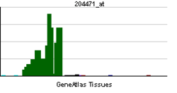
| RNA expression pattern | |||||||||||||
 | |||||||||||||
 | |||||||||||||
| More reference expression data | |||||||||||||
| Orthologs | |||||||||||||
| Species | Human | Mouse | |||||||||||
| Entrez | 2596 | 14432 | |||||||||||
| Ensembl | ENSG00000172020 | ENSMUSG00000047261 | |||||||||||
| UniProt | P17677 | P06837 | |||||||||||
| RefSeq (mRNA) | NM_001130064 | NM_008083 | |||||||||||
| RefSeq (protein) | NP_001123536 | NP_032109 | |||||||||||
| Location (UCSC) | Chr 3: 115.34 – 115.44 Mb | Chr 16: 42.25 – 42.34 Mb | |||||||||||
| PubMed search | |||||||||||||
Growth Associated Protein 43 also known as GAP43 is a protein that in humans is encoded by the GAP43 gene.[1]
GAP43 has been termed a 'growth' or 'plasticity' protein because it is expressed at high levels in neuronal growth cones during development, during axonal regeneration and is phosphorylated after long-term potentiation (LTP) and after learning (reference needed). This protein is considered a crucial component of the axon and presynaptic terminal, its null mutation leading to death within days after birth due to axon pathfinding defects.[2]
Synonyms
GAP43 is also referred to as:
- protein F1
- neuromodulin
- neural phosphoprotein B-50
- axonal membrane protein GAP-43
- calmodulin-binding protein P-57
- nerve growth-related peptide GAP43
- neuron growth-associated protein 43
Function
GAP43, is a nervous tissue-specific cytoplasmic protein that can be attached to the membrane via a dual palmitoylation sequence on cysteines 3 and 4. This sequence targets GAP43 to lipid rafts. It is a major protein kinase C (PKC) substrate and is considered to play a key role in neurite formation, regeneration, and plasticity.[3][4] The role of GAP-43 in CNS development is not limited to effects on axons: It is also a component of the centrosome, and differentiating neurons that do not express GAP-43 show mislocalization of the centrosome and mitotic spindles, particularly in neurogenic cell divisions. As a consequence, in the cerebellum, the neuronal precursor pool fails to expand normally and the cerebellum is significantly smaller.
Several different laboratories studying the same protein, now called GAP43, initially used different names. It was designated F1, then B-50, then GAP43, pp46, and finally neuromodulin, each name reflecting a different function of the same molecule.[5] F1 was localized to synapses, and was increased in its phosphorylation one day after learning. However, F1 was not cAMP kinase dependent. B-50 was regulated by the pituitary peptide ACTH and was associated with grooming behavior. In the case of GAP-43, it was designated as a growth-associated protein because its synthesis was upregulated during axonal regeneration. Pp46 was concentrated in neuronal growth cones and was thus postulated to play an important role in brain development. In the case of neuromodulin, it was shown to bind calmodulin avidly.
GAP43, the consensus choice for its designation,[5] is a nervous system-specific protein that is attached to the membrane via a dual palmitoylation sequence on cysteines 3 and 4, though it can exist in the non-bound form in the cytoplasm. This dual sequence enables the association of phosphatidylinositol-4,5-bisphosphate [PI(4,5)P2] or PIP2, with actin, facilitating the latter’s polymerization thereby regulating neuronal structure. This can occur within a lipid raft so as to compartmentalize and localize motility of filopodia in growth cones in developing brains, and could also remodel presynaptic terminals in adults in an activity-dependent manner. GAP-43 is also a protein kinase C (PKC) substrate. Phosphorylation of serine-41 on GAP-43 by PKC regulates neurite formation, regeneration, and synaptic plasticity.[3]
Because of the association and potential binding of GAP43 with a number of different molecules, including PKC, PIP2, actin, calmodulin, spectrin, palmitate, synaptophysin, amyloid and tau protein, it may be useful to think of GAP43 as an adaptor protein situated within the terminal in a supramolecular complex regulating presynaptic terminal functions, particularly bidirectional communication with the postsynaptic process. Its important role in memory and information storage is executed through its cell biological mechanisms of phosphorylation, palmitoylation, protein-protein interaction and structural remodeling via actin polymerization.
Clinical significance
Humans with a deletion in one allele of the GAP43 gene fail to form telencephalic commissures and are mentally retarded,.[6][7]
Model organisms
| Characteristic | Phenotype |
|---|---|
| Homozygote viability | Abnormal |
| Recessive lethal study | Normal |
| Fertility | Normal |
| Body weight | Normal |
| Anxiety | Normal |
| Neurological assessment | Normal |
| Grip strength | Normal |
| Hot plate | Normal |
| Dysmorphology | Normal |
| Indirect calorimetry | Normal |
| Glucose tolerance test | Normal |
| Auditory brainstem response | Normal |
| DEXA | Normal |
| Radiography | Normal |
| Body temperature | Normal |
| Eye morphology | Normal |
| Clinical chemistry | Normal |
| Plasma immunoglobulins | Abnormal |
| Haematology | Normal |
| Micronucleus test | Normal |
| Heart weight | Normal |
| Brain histopathology | Normal |
| Eye Histopathology | Normal |
| Salmonella infection | Normal[8] |
| Citrobacter infection | Normal[9] |
| All tests and analysis from[10][11] |
Model organisms have been used in the study of GAP43 function. A conditional knockout mouse line, called Gap43tm1a(EUCOMM)Wtsi[12][13] was generated as part of the International Knockout Mouse Consortium program — a high-throughput mutagenesis project to generate and distribute animal models of disease to interested scientists.[14][15][16] Male and female animals underwent a standardized phenotypic screen to determine the effects of deletion.[10][17] Twenty five tests were carried out on mutant mice and two significant abnormalities were observed. No homozygous mutant mice survived until weaning. The remaining tests were carried out on heterozygous mutant adult mice and increased IgG1 levels were observed in these animals.[10]
Studies on another homozygous GAP43 knockout mouse line found it to be lethal days after birth because it plays a critical role in the development of the mammalian CNS.[18] Telencephalic commissures fail to form,[19] thalamocortical afferents are mistargeted, especially in somatosensory, particularly barrel, cortex.[18] GAP43 is not only important for axon targeting during development but it has been shown to be important also for the maintenance of the structure and dynamics of axonal fibres and of their synaptic terminals in wild-type rodents both during normal conditions and during lesion-induced axonal sprouting.[20][21][22] The cerebellum is also affected.[23] GAP43 is also haploinsufficient for the cortical phenotypes and the severity of the axon targeting phenotype is directly related to the extent to which the affected axons are phosphorylated by PKC, suggesting that axons require a functional threshold of phosphorylated GAP43 for targeting to occur normally.[19] Moreover, elevation above this threshold in GAP43 mice can enhance learning and also facilitate a physiological model of learning, long-term potentiation (LTP).[24] However, further enrichment beyond a certain level can be devastating to cognitive functions.
References
- ↑ Kosik KS, Orecchio LD, Bruns GA, Benowitz LI, MacDonald GP, Cox DR, Neve RL (April 1988). "Human GAP-43: its deduced amino acid sequence and chromosomal localization in mouse and human". Neuron 1 (2): 127–32. doi:10.1016/0896-6273(88)90196-1. PMID 3272162.
- ↑ "Entrez Gene: GAP43 growth associated protein 43".
- ↑ 3.0 3.1 Benowitz LI, Routtenberg A (February 1997). "GAP-43: an intrinsic determinant of neuronal development and plasticity". Trends Neurosci. 20 (2): 84–91. doi:10.1016/S0166-2236(96)10072-2. PMID 9023877.
- ↑ Aarts LH, Schotman P, Verhaagen J, Schrama LH, Gispen WH (1998). "The role of the neural growth associated protein B-50/GAP-43 in morphogenesis". Adv. Exp. Med. Biol. 446: 85–106. doi:10.1007/978-1-4615-4869-0_6. PMID 10079839.
- ↑ 5.0 5.1 Benowitz LI, Routtenberg A (1987). "A membrane phosphoprotein associated with neural development, axonal regeneration, phospholipid metabolism, and synaptic plasticity". Trends in Neurosciences 10 (12): 527–532. doi:10.1016/0166-2236(87)90135-4.
- ↑ Genuardi M, Calvieri F, Tozzi C, Coslovi R, Neri G (Oct 1994). "A new case of interstitial deletion of chromosome 3q, del(3q)(q13.12q21.3), with agenesis of the corpus callosum.". Clin Dysmorphol 3 (4): 292–6. doi:10.1097/00019605-199410000-00003. PMID 7894733.
- ↑ Mackie Ogilvie C, Rooney SC, Hodgson SV, Berry AC (Mar 1998). "Deletion of chromosome 3q proximal region gives rise to a variable phenotype". Clin Genet 53 (3): 220–2. doi:10.1111/j.1399-0004.1998.tb02681.x. PMID 9630079.
- ↑ "Salmonella infection data for Gap43". Wellcome Trust Sanger Institute.
- ↑ "Citrobacter infection data for Gap43". Wellcome Trust Sanger Institute.
- ↑ 10.0 10.1 10.2 Gerdin AK (2010). "The Sanger Mouse Genetics Programme: High throughput characterisation of knockout mice". Acta Ophthalmologica 88: 925–7. doi:10.1111/j.1755-3768.2010.4142.x.
- ↑ Mouse Resources Portal, Wellcome Trust Sanger Institute.
- ↑ "International Knockout Mouse Consortium".
- ↑ "Mouse Genome Informatics".
- ↑ Skarnes, W. C.; Rosen, B.; West, A. P.; Koutsourakis, M.; Bushell, W.; Iyer, V.; Mujica, A. O.; Thomas, M.; Harrow, J.; Cox, T.; Jackson, D.; Severin, J.; Biggs, P.; Fu, J.; Nefedov, M.; De Jong, P. J.; Stewart, A. F.; Bradley, A. (2011). "A conditional knockout resource for the genome-wide study of mouse gene function". Nature 474 (7351): 337–342. doi:10.1038/nature10163. PMC 3572410. PMID 21677750.
- ↑ Dolgin E (2011). "Mouse library set to be knockout". Nature 474 (7351): 262–3. doi:10.1038/474262a. PMID 21677718.
- ↑ Collins FS, Rossant J, Wurst W (2007). "A Mouse for All Reasons". Cell 128 (1): 9–13. doi:10.1016/j.cell.2006.12.018. PMID 17218247.
- ↑ van der Weyden L, White JK, Adams DJ, Logan DW (2011). "The mouse genetics toolkit: revealing function and mechanism.". Genome Biol 12 (6): 224. doi:10.1186/gb-2011-12-6-224. PMC 3218837. PMID 21722353.
- ↑ 18.0 18.1 Strittmatter SM, Fankhauser C, Huang PL, Mashimo H, Fishman MC (February 1995). "Neuronal pathfinding is abnormal in mice lacking the neuronal growth cone protein GAP-43". Cell 80 (3): 445–52. doi:10.1016/0092-8674(95)90495-6. PMID 7859286.
- ↑ 19.0 19.1 Shen Y, Mani S, Donovan SL, Schwob JE, Meiri KF (January 2002). "Growth-associated protein-43 is required for commissural axon guidance in the developing vertebrate nervous system". J. Neurosci. 22 (1): 239–47. PMID 11756507.
- ↑ Grasselli G, Mandolesi G, Strata P, Cesare P (June 2011). "Impaired Sprouting and Axonal Atrophy in Cerebellar Climbing Fibres following In Vivo Silencing of the Growth-Associated Protein GAP43". PLoS ONE 6 (6): e20791. doi:10.1371/journal.pone.0020791. PMID 21695168.
- ↑ Grasselli G, Strata P (February 2013). "Structural plasticity of climbing fibers and the growth-associated protein GAP-43". Front. Neural Circuits 7 (25). doi:10.3389/fncir.2013.00025. PMID 23441024.
- ↑ Allegra Mascaro AL, Cesare P, Sacconi L, Grasselli G, Mandolesi G, Maco B, Knott G, Huag L, De Paola V, Strata P and Pavone FS (2013). "In vivo single branch axotomy induces GAP-43 dependent sprouting and synaptic remodeling in cerebellar cortex". Proc Natl Acad Sci U S A 110 (26): 10824–9. doi:10.1073/pnas.1219256110. PMID 23754371.
- ↑ Shen Y, Mishra R, Mani S, Meiri KF (2008). "Both cell-autonomous and cell non-autonomous functions of GAP43 are required for normal patterning of the cerebellum in vivo". Cerebellum 7 (3): 451–66. doi:10.1007/s12311-008-0049-5. PMID 18777197.
- ↑ Routtenberg A, Cantallops I, Zaffuto S, Serrano P, Namgung U (June 2000). "Enhanced learning after genetic overexpression of a brain growth protein". Proc. Natl. Acad. Sci. U.S.A. 97 (13): 7657–62. doi:10.1073/pnas.97.13.7657. PMC 16601. PMID 10861025.
Further reading
- Fantini F, Johansson O (1993). "Expression of growth-associated protein 43 and nerve growth factor receptor in human skin: a comparative immunohistochemical investigation.". J. Invest. Dermatol. 99 (6): 734–42. doi:10.1111/1523-1747.ep12614465. PMID 1281863.
- Mercken M, Lübke U, Vandermeeren M et al. (1992). "Immunocytochemical detection of the growth-associated protein B-50 by newly characterized monoclonal antibodies in human brain and muscle.". J. Neurobiol. 23 (3): 309–21. doi:10.1002/neu.480230310. PMID 1385623.
- Spencer SA, Schuh SM, Liu WS, Willard MB (1992). "GAP-43, a protein associated with axon growth, is phosphorylated at three sites in cultured neurons and rat brain.". J. Biol. Chem. 267 (13): 9059–64. PMID 1533624.
- Apel ED, Litchfield DW, Clark RH et al. (1991). "Phosphorylation of neuromodulin (GAP-43) by casein kinase II. Identification of phosphorylation sites and regulation by calmodulin.". J. Biol. Chem. 266 (16): 10544–51. PMID 1828073.
- Apel ED, Byford MF, Au D et al. (1990). "Identification of the protein kinase C phosphorylation site in neuromodulin.". Biochemistry 29 (9): 2330–5. doi:10.1021/bi00461a017. PMID 2140056.
- Kosik KS, Orecchio LD, Bruns GA et al. (1990). "Human GAP-43: its deduced amino acid sequence and chromosomal localization in mouse and human.". Neuron 1 (2): 127–32. doi:10.1016/0896-6273(88)90196-1. PMID 3272162.
- Ng SC, de la Monte SM, Conboy GL et al. (1990). "Cloning of human GAP-43: growth association and ischemic resurgence.". Neuron 1 (2): 133–9. doi:10.1016/0896-6273(88)90197-3. PMID 3272163.
- Nielander HB, De Groen PC, Eggen BJ et al. (1993). "Structure of the human gene for the neural phosphoprotein B-50 (GAP-43).". Brain Res. Mol. Brain Res. 19 (4): 293–302. doi:10.1016/0169-328X(93)90128-C. PMID 8231732.
- Oehrlein SA, Parker PJ, Herget T (1996). "Phosphorylation of GAP-43 (growth-associated protein of 43 kDa) by conventional, novel and atypical isotypes of the protein kinase C gene family: differences between oligopeptide and polypeptide phosphorylation.". Biochem. J. 317. ( Pt 1): 219–24. PMC 1217466. PMID 8694767.
- Kanazir S, Ruzdijic S, Vukosavic S et al. (1997). "GAP-43 mRNA expression in early development of human nervous system.". Brain Res. Mol. Brain Res. 38 (1): 145–55. doi:10.1016/0169-328X(96)00008-3. PMID 8737678.
- de Groen PC, Eggen BJ, Gispen WH et al. (1996). "Cloning and promoter analysis of the human B-50/GAP-43 gene.". J. Mol. Neurosci. 6 (2): 109–19. doi:10.1007/BF02736770. PMID 8746449.
- Chao S, Benowitz LI, Krainc D, Irwin N (1997). "Use of a two-hybrid system to investigate molecular interactions of GAP-43.". Brain Res. Mol. Brain Res. 40 (2): 195–202. doi:10.1016/0169-328X(96)00049-6. PMID 8872303.
- Gamby C, Waage MC, Allen RG, Baizer L (1996). "Analysis of the role of calmodulin binding and sequestration in neuromodulin (GAP-43) function.". J. Biol. Chem. 271 (43): 26698–705. doi:10.1074/jbc.271.43.26698. PMID 8900147.
- Heuss D, Schlötzer-Schrehardt U (1998). "Subcellular localization of phosphoprotein B-50 in regenerating muscle. An immuno-electron microscopic study.". Neurol. Res. 20 (4): 360–4. PMID 9618702.
- Neve RL, Coopersmith R, McPhie DL et al. (1998). "The neuronal growth-associated protein GAP-43 interacts with rabaptin-5 and participates in endocytosis.". J. Neurosci. 18 (19): 7757–67. PMID 9742146.
- Arni S, Keilbaugh SA, Ostermeyer AG, Brown DA (1998). "Association of GAP-43 with detergent-resistant membranes requires two palmitoylated cysteine residues.". J. Biol. Chem. 273 (43): 28478–85. doi:10.1074/jbc.273.43.28478. PMID 9774477.
- Eastwood SL, Harrison PJ (1999). "Hippocampal and cortical growth-associated protein-43 messenger RNA in schizophrenia.". Neuroscience 86 (2): 437–48. doi:10.1016/S0306-4522(98)00040-2. PMID 9881859.
- Cargill M, Altshuler D, Ireland J et al. (1999). "Characterization of single-nucleotide polymorphisms in coding regions of human genes.". Nat. Genet. 22 (3): 231–8. doi:10.1038/10290. PMID 10391209.
- Riederer BM, Routtenberg A (1999). "Can GAP-43 interact with brain spectrin?". Brain Res. Mol. Brain Res. 71 (2): 345–8. doi:10.1016/S0169-328X(99)00179-5. PMID 10521589.
- Vento P, Soinila S (1999). "Quantitative comparison of growth-associated protein GAP-43, neuron-specific enolase, and protein gene product 9.5 as neuronal markers in mature human intestine.". J. Histochem. Cytochem. 47 (11): 1405–16. doi:10.1177/002215549904701107. PMID 10544214.
External links
- Gap-43 protein at the US National Library of Medicine Medical Subject Headings (MeSH)
- ihop-net, Growth associated protein 43
- NCBI
| ||||||||||||||||||||||||||||||||||||||||||