Fourth ventricle
| Fourth ventricle | |
|---|---|
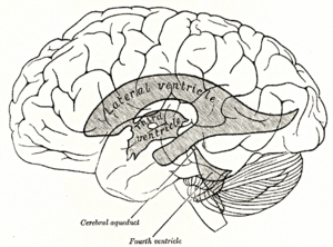 Scheme showing relations of the ventricles to the surface of the brain. (Fourth ventricle labeled at bottom center.) | |
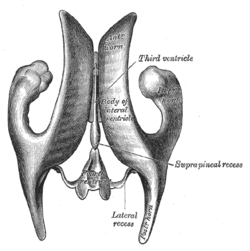 Drawing of a cast of the ventricular cavities, viewed from above. (Fourth ventricle visible at bottom center.) | |
| Details | |
| Latin | ventriculus quartus |
| Identifiers | |
| Gray's | p.797 |
| MeSH | A08.186.211.276.500 |
| NeuroNames | hier-617 |
| NeuroLex ID | Fourth ventricle |
| TA | A14.1.05.701 |
| FMA | 78469 |
| Anatomical terms of neuroanatomy | |
The fourth ventricle is one of the four connected fluid-filled cavities within the human brain. These cavities, known collectively as the ventricular system, consist of the left and right lateral ventricles, the third ventricle, and the fourth ventricle. The fourth ventricle extends from the cerebral aqueduct (aqueduct of Sylvius) to the obex, and is filled with cerebrospinal fluid (CSF).
The fourth ventricle has a characteristic diamond shape in cross-sections of the human brain. It is located within the pons or in the upper part of the medulla. CSF entering the fourth ventricle through the cerebral aqueduct can exit to the subarachnoid space of the spinal cord through two lateral foramina of Luschka (singular: foramen of Luschka) and a single, midline foramen of Magendie (see List of human anatomical parts named after people).
Roof and floor
The fourth ventricle has a "roof" dorsally and a "floor" ventrally. The roof of the fourth ventricle is formed by the cerebellum (superior and inferior medullary vela), the floor by the rhomboid fossa, and the side "walls" formed by the cerebellar peduncles. Among the prominent features of the floor of the fourth ventricle are the:
- facial colliculus: formed by the internal part of the facial nerve as it loops around the abducens nucleus in the lower pons;
- sulcus limitans: which represents the border between the alar plate and the basal plate of the developing neural tube;
- obex: represents the caudal tip of the fourth ventricle; the obex is also a marker for the level of the foramen magnum of the skull and therefore is a marker for the imaginary dividing line between the medulla and spinal cord.
- "median sulcus" - divides the floor into right and left halves. It extends from cerebral aqueduct of the midbrain to central canal of the spinal cord.
- "stria medullaris" - fibers derived from arcuate nuclei, which emerge from the median sulcus and run transversely across the floor to enter into the inferior cerebellar penducle.
- "medial eminence" - elevations on either side of the median sulcus.
- "sulcus limitans" - medial eminence is laterally bounded by sulcus limitans.
- "vestibular area" - lateral to sulcus limitans vestibular nuclei is overlied by this.
- The upper end of the sulcus limitans widens into a triangular depression called "superior fovea". Above the superior fovea sulcus limitans presents a flattened grey area called "locus ceruleus".
- The lower end of the sulcus limitans widens into a triangular depression called "Inferior fovea".
- Other features are the hypoglossal trigone and the vagal trigone.
Development
The fourth ventricle, similarly to other parts of the ventricular system of the brain, develops from the central canal of the neural tube. Specifically, the fourth ventricle originates from the portion of the tube that is present in the developing rhombencephalon.[1] During the first trimester of pregnancy central canal expands into lateral, third and fourth ventricles, connected by thinner channels.[2] In lateral ventricles specialized areas- choroid plexuses appear, which produce cerebrospinal fluid. If its production is bigger than resorption or its circulation is blocked- the enlargement of the ventricles may appear and cause a hydrocephalus. Fetal lateral ventricles may be diagnosed using linear or planar measurements.[3] See also Dandy-Walker syndrome.
Clinical significance
The fourth ventricle is a common location of an intracranial ependymomal tumour.
References
- ↑ Le, Tao; Bhushan, Vikas; Vasan, Neil (2010). First Aid for the USMLE Step 1: 2010 20th Anniversary Edition. USA: The McGraw-Hill Companies, Inc. p. 126. ISBN 978-0-07-163340-6.
- ↑ Carlson, Bruce M. (1999). Human Embryology & Developmental Biology. Mosby. pp. 237–238. ISBN 0-8151-1458-3.
- ↑ Glonek, M; Kedzia, A; Derkowski, W (2003). "Planar measurements of foetal lateral ventricles.". Folia morphologica 62 (3): 263–5. PMID 14507062.
Additional images
-

Transverse section of medulla oblongata below the middle of the olive.
-
Diagram showing the course of the arcuate fibers.
-

The formatio reticularis of the medulla oblongata, shown by a transverse section passing through the middle of the olive.
-

Coronal section of the pons, at its upper part.
-
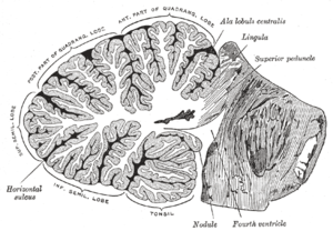
Sagittal section of the cerebellum, near the junction of the vermis with the hemisphere.
-
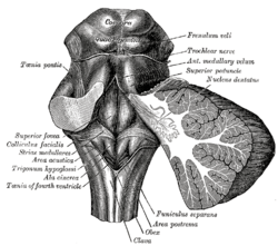
Rhomboid fossa.
-

Mesal aspect of a brain sectioned in the median sagittal plane.
-
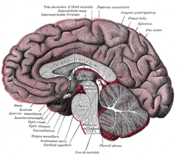
Median sagittal section of brain.
-

Drawing of a cast of the ventricular cavities, viewed from the side.
-
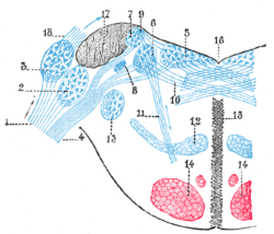
Terminal nuclei of the vestibular nerve, with their upper connections.
-
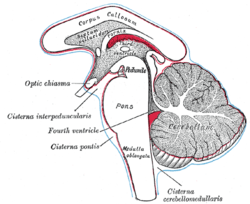
Diagram showing the positions of the three principal subarachnoid cisternæ.
-
Third ventricle is the vertical dark slit in the middle of the field.
-

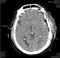
Fourth ventricle
External links
| Wikimedia Commons has media related to Fourth ventricle. |
- Atlas image: n2a8p1 at the University of Michigan Health System - "Fourth Ventricle, Sagittal Section, Medial View"
- Stained brain slice images which include the "fourth%20ventricle" at the BrainMaps project
- fourth+ventricle at eMedicine Dictionary
- Anatomy diagram: 13048.000-3 at Roche Lexicon - illustrated navigator, Elsevier
| ||||||||||||||||||||||||||||||||||||||||||||||||||||