Focal nodular hyperplasia
| Focal nodular hyperplasia | |
|---|---|
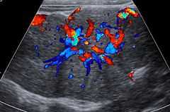
 Micrograph of focal nodular hyperplasia. H&E stain. | |
| Classification and external resources | |
| DiseasesDB | 33467 |
| eMedicine | radio/286 |
| MeSH | D020518 |
Focal nodular hyperplasia (FNH) is a benign tumor of the liver (hepatic tumor), which is the second most prevalent tumor of the liver (the first is hepatic hemangioma). It is usually asymptomatic, rarely grows or bleeds, and has no malignant potential. This tumour was once often resected because it was difficult to distinguish from hepatic adenoma, but with modern multiphase imaging is usually now diagnosed by strict imaging criteria and not resected.
Presentation
Focal nodular hyperplasia's most recognizable gross feature is a central stellate scar seen in 60–70% of cases. Microscopically, a lobular proliferation of bland-appearing hepatocytes with a bile ductular proliferation and malformed vessels within the fibrous scar is the most common pattern. Other patterns include telangiectatic, hyperplastic-adenomatous, and lesions with focal large-cell dysplasia.[1] Rarely, these lesions may be multiple or can occur as part of a syndrome with hemangiomas, epithelioid hemangioendothelioma, hepatic adenomas, fibrolamellar hepatocellular carcinoma, vascular malformations of the brain, meningiomas, and/or astrocytomas.[1]
Notes
- ↑ 1.0 1.1 Nguyen BN, Flejou JF, Terris B, Belghiti J, Degott C (December 1999). "Focal nodular hyperplasia of the liver: a comprehensive pathologic study of 305 lesions and recognition of new histologic forms". Am. J. Surg. Pathol. 23 (12): 1441–54. doi:10.1097/00000478-199912000-00001. PMID 10584697.
References
- Hsee LC, McCall JL, Koea JB (2005). "Focal nodular hyperplasia: what are the indications for resection?". HPB (Oxford) 7 (4): 298–302. doi:10.1080/13651820500273624. PMC 2043107. PMID 18333211.
| ||||||||||||||||||||||||||||||||||||||||||||||||||||||||||||||||||||||||