Fibula
| Fibula | |
|---|---|
 Position of fibula in human (shown in red) | |
 Cross section of human lower leg, showing fibula in centre (latin terminology) | |
| Details | |
| Latin | (os) fibula |
| Articulations | Superior and inferior tibiofibular joint |
| Identifiers | |
| Gray's | p.260 |
| MeSH | A02.835.232.043.650.321 |
| TA | A02.5.07.001 |
| FMA | 24479 |
| Anatomical terms of bone | |
The fibula (/ˈfɪbjʉlə/[1][2]) or calf bone is a leg bone located on the lateral side of the tibia, with which it is connected above and below. It is the smaller of the two bones, and, in proportion to its length, the slenderest of all the long bones. Its upper extremity is small, placed toward the back of the head of the tibia, below the level of the knee joint, and excluded from the formation of this joint. Its lower extremity inclines a little forward, so as to be on a plane anterior to that of the upper end; it projects below the tibia, and forms the lateral part of the ankle joint.
Structure
The bone has the following components:
- Lateral malleolus
- Interosseous membrane connecting the fibula to the tibia, forming a syndesmoses joint
- The superior tibiofibular articulation is an arthrodial joint between the lateral condyle of the tibia and the head of the fibula.
- The inferior tibiofibular articulation (tibiofibular syndesmosis) is formed by the rough, convex surface of the medial side of the lower end of the fibula, and a rough concave surface on the lateral side of the tibia.
Blood supply
The blood supply is important for planning free tissue transfer because the fibula is commonly used to reconstruct the mandible. The shaft is supplied in its middle third by a large nutrient vessel from the fibular artery. It is also perfused from its periosteum which receives many small branches from the fibular artery. The proximal head and the epiphysis are supplied by a branch of the anterior tibial artery. In harvesting the bone the middle third is always taken and the ends preserved (4 cm proximally and 6 cm distally)
Development
The fibula is ossified from three centers, one for the shaft, and one for either end. Ossification begins in the body about the eighth week of fetal life, and extends toward the extremities. At birth the ends are cartilaginous.
Ossification commences in the lower end in the second year, and in the upper about the fourth year. The lower epiphysis, the first to ossify, unites with the body about the twentieth year; the upper epiphysis joins about the twenty-fifth year.
Function
The fibula does not carry any significant load (weight) of the body. It extends past the lower end of the tibia and forms the outer part of the ankle providing stability to this joint. It has grooves for certain ligaments which gives them leverage and multiplies the muscle force. It provides attachment points for the following muscles:
 Muscle attachments (seen from the front) |  Muscle attachments (seen from the back) |
| Muscle | Direction | Attachment[3] |
| Biceps femoris muscle | Insertion | Head of fibula |
| Extensor hallucis longus muscle | Origin | Medial side of fibula |
| Extensor digitorum longus muscle | Origin | Proximal part of the medial side of fibulua |
| Fibularis tertius | Origin | Distal part of the medial side of fibulua |
| Fibularis longus | Origin | Head and the lateral side of fibula |
| Fibularis brevis | Origin | Distal 2/3 of the lateral side of fibula |
| Soleus muscle | Origin | Proximal 1/3 of the posterior side of fibula |
| Tibialis posterior muscle | Origin | Lateral part of the posterior side of fibula |
| Flexor hallucis longus muscle | Origin | Posterior side of fibula |
Clinical significance
Avulsion fracture
An avulsion fracture of the head of the fibula refers to the fracture of the fibular head because of a sudden contraction of the biceps femoris muscle that pulls its site of attachment on the bone. The attachment of the biceps femoris tendon on the fibular head is closely related to the lateral collateral ligament of the knee. Therefore, this ligament is prone to injury in this type of avulsion fracture.[4]
History
Etymology
The word fibula can be dated back to c. 1670 to describe a clasp or brooch – see fibula (brooch) – and was first used in English for the smaller bone in the lower leg c. 1706. It derives from Latin fībula, also meaning a clasp or brooch. The bone was so called because it resembles a clasp like a modern safety pin.[5]
In other animals
Because the fibula bears relatively little weight in comparison with the tibia, it is typically narrower in all but the most primitive tetrapods. In many animals, it still articulates with the posterior part of the lower extremity of the femur, but this feature is frequently lost (as it is in humans). In some animals, the reduction of the fibula has proceeded even further than it has in humans, with the loss of the tarsal articulation, and, in extreme cases (such as the horse), partial fusion with the tibia.[6]
See also
- This article uses anatomical terminology; for an overview, see anatomical terminology.
- Peroneal
References
This article incorporates text in the public domain from the 20th edition of Gray's Anatomy (1918)
- ↑ OED 2nd edition, 1989.
- ↑ Entry "fibula" in Merriam-Webster Online Dictionary.
- ↑ Bojsen-Møller, Finn; Simonsen, Erik B.; Tranum-Jensen, Jørgen (2001). Bevægeapparatets anatomi [Anatomy of the Locomotive Apparatus] (in Danish) (12th ed.). pp. 364–367. ISBN 978-87-628-0307-7.
- ↑ Gottsegen, CJ; Eyer, BA; White, EA; Learch, TJ; Forrester, D (2008). "Avulsion fractures of the knee: imaging findings and clinical significance.". Radiographics 28 (6): 1755–1770. doi:10.1148/rg.286085503. PMID 18936034.
- ↑ etymonline.com
- ↑ Romer, Alfred Sherwood; Parsons, Thomas S. (1977). The Vertebrate Body. Philadelphia, PA: Holt-Saunders International. p. 205. ISBN 0-03-910284-X.
Additional Images
-

Position of fibula (shown in red)
-

Shape of fibula (right)
-
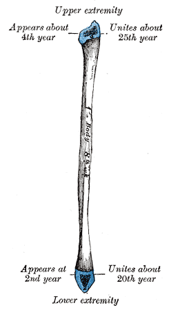
Diagram which depicts ossification of human fibula.
-

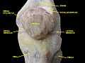
Lower extremity of right fibula. Medial aspect.
-

Ankle joint. Deep dissection. Lateral view.
-
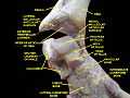
Ankle joint. Deep dissection.
-

Ankle joint. Deep dissection.
-
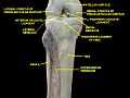
Knee and tibiofibular joint.Deep dissection. Anterior view.
-

Knee and tibiofibular joint.Deep dissection. Anterior view.
-
Knee joint.Deep dissection. Anteromedial view.
-

Knee, tibiofibular and ankle joints.Deep dissection. Anterolateral view.
-

Knee, tibiofibular and ankle joints.Deep dissection. Anterolateral view.
-

Ankle joint. Bones of foot.Deep dissection.
-

Knee joint. Deep dissection. Posterior view.
External links
| Wikimedia Commons has media related to Fibula (bone). |
| Look up fibula in Wiktionary, the free dictionary. |
- Anatomy photo:17:st-1402 at the SUNY Downstate Medical Center
- Osteocutaneous fibula flap video on Youtube.com
| ||||||||||||||||||||||||||||||||||||||||||||||||||||||||||||||||||||||