Ferritin
| Ferritin | |||||||||
|---|---|---|---|---|---|---|---|---|---|
 | |||||||||
| Identifiers | |||||||||
| Pfam | PF00210 | ||||||||
| Pfam clan | CL0044 | ||||||||
| InterPro | IPR008331 | ||||||||
| SCOP | 1fha | ||||||||
| SUPERFAMILY | 1fha | ||||||||
| |||||||||
| ferritin, light polypeptide | |
|---|---|
| Identifiers | |
| Symbol | FTL |
| Entrez | 2512 |
| HUGO | 3999 |
| OMIM | 134790 |
| RefSeq | NM_000146 |
| UniProt | P02792 |
| Other data | |
| Locus | Chr. 19 q13.3–13.4 |
| ferritin, heavy polypeptide 1 | |
|---|---|
| Identifiers | |
| Symbol | FTH1 |
| Alt. symbols | FTHL6 |
| Entrez | 2495 |
| HUGO | 3976 |
| OMIM | 134770 |
| RefSeq | NM_002032 |
| UniProt | P02794 |
| Other data | |
| Locus | Chr. 11 q13 |
| ferritin mitochondrial | |
|---|---|
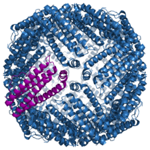
 Crystallographic structure of mitochondrial ferritin.[2] | |
| Identifiers | |
| Symbol | FTMT |
| Entrez | 94033 |
| HUGO | 17345 |
| OMIM | 608847 |
| RefSeq | NM_177478 |
| UniProt | Q8N4E7 |
| Other data | |
| Locus | Chr. 5 q23.1 |
Ferritin is a ubiquitous intracellular protein that stores iron and releases it in a controlled fashion. The protein is produced by almost all living organisms, including algae, bacteria, higher plants, and animals. In humans, it acts as a buffer against iron deficiency and iron overload.[3] Ferritin is found in most tissues as a cytosolic protein, but small amounts are secreted into the serum where it functions as an iron carrier. Plasma ferritin is also an indirect marker of the total amount of iron stored in the body, hence serum ferritin is used as a diagnostic test for iron deficiency anemia.[4]
Ferritin is a globular protein complex consisting of 24 protein subunits and is the primary intracellular iron-storage protein in both prokaryotes and eukaryotes, keeping iron in a soluble and non-toxic form. Ferritin that is not combined with iron is called apoferritin.
Gene
Ferritin genes are highly conserved between species. All vertebrate ferritin genes have three introns and four exons.[5] In human ferritin, introns are present between the 34/5th, 82/3rd, and 14/5th amino acid residues; in addition, there are one to two hundred untranslated bases at either end of the combined exons.[6] The tyrosine residue at amino acid position 27 is thought to be associated with biomineralization.[7]
Protein structure
Ferritin is a protein of 450 kDa consisting of 24 subunits that is present in every cell type.[6] In vertebrates, these subunits are both the light (L) and the heavy (H) type with an apparent molecular weight of 19 kDa or 21 kDa respectively; their sequences are about 50% homologous.[6] Amphibians have an additional ("M") type of ferritin;[8] the single ferritin of plants and bacteria most closely resembles the vertebrate H-type.[8] Two types have been recovered in the gastropod Lymnaea, the somatic ferritin being distinct from the yolk ferritin (see below).[8] An additional subunit resembling Lymnaea soma ferritin is associated with shell formation in the pearl oyster.[9] Two types are present in the parasite Schistosoma, one in males, the other in females.[8] All the aforementioned ferritins are similar, in terms of their primary sequence, with the vertebrate H-type.[8] In E. coli, a 20% similarity to human H-ferritin is observed.[8] Inside the ferritin shell, iron ions form crystallites together with phosphate and hydroxide ions. The resulting particle is similar to the mineral ferrihydrite. Each ferritin complex can store about 4500 iron (Fe3+) ions.[6][8]
Some ferritin complexes in vertebrates are hetero-oligomers of two highly related gene products with slightly different physiological properties. The ratio of the two homologous proteins in the complex depends on the relative expression levels of the two genes.
Mitochondrial ferritin was recently identified as a protein precursor, and is classified as a metal-binding protein that is located within the mitochondria.[10] After the protein is taken up by the mitochondria it can be processed into a mature protein and assemble to form functional ferritin shells. Its structure was determined at 1.70 angstroms through the use of X-ray diffraction and contains 182 residues. It is 67% helical. The Ramachandran plot [11] shows that the structure of mitochondrial ferritin is mainly alpha helical with a low prevalence of beta sheets. Unlike other human ferritin, it appears to have no introns in its genetic code.
Function
Iron storage
Ferritin serves to store iron in a non-toxic form, to deposit it in a safe form, and to transport it to areas where it is required.[12] The function and structure of the expressed ferritin protein varies in different cell types. This is controlled primarily by the amount and stability of mRNA. mRNA concentration is further tweaked by changes to how it is stored and how efficiently it is transcribed.[6] The presence of iron itself is a major trigger for the production of ferritin,[6] with some exceptions (such as the yolk ferritin of the gastropod Lymnaea, which lacks an iron-responsive unit).[8]
Free iron is toxic to cells as it acts as a catalyst in the formation of free radicals from reactive oxygen species via the Fenton Reaction.[13] Hence vertebrates evolve an elaborate set of protective mechanisms to bind iron in various tissue compartments. Within cells, iron is stored in a protein complex as ferritin or hemosiderin. Apoferritin binds to free ferrous iron and stores it in the ferric state. As ferritin accumulates within cells of the reticuloendothelial system, protein aggregates are formed as hemosiderin. Iron in ferritin or hemosiderin can be extracted for release by the RE cells although hemosiderin is less readily available. Under steady state conditions, the serum ferritin level correlates with total body iron stores; thus, the serum ferritin FR5Rl is the most convenient laboratory test to estimate iron stores.
Because iron is an important mineral in mineralization, ferritin is employed in the shells of organisms such as molluscs to control the concentration and distribution of iron, thus sculpting shell morphology and colouration.[14][15] It also plays a role in the haemolymph of the polyplacophora where it serves to rapidly transport iron to the mineralizing radula.[16]
Ferroxidase activity
Vertebrate ferritin consists of two or three subunits which are named based on their molecular weight: L "light", H "heavy", and M "middle" subunits. M subunit has only been reported in bullfrog. In bacteria and archaea ferritin consists of one subunit type.[17] H and M subunits of eukaryotic ferritin and all subunits of bacterial and archaeal ferritin are H-type and have ferroxidase activity, which is conversion of iron from the ferrous (Fe2+) to ferric (Fe3+) forms. This limits the deleterious reaction which occurs between ferrous iron and hydrogen peroxide known as the Fenton reaction which produces the highly damaging hydroxyl radical. The ferroxidase activity occurs at a diiron binding site in the middle of each H-type subunits.[17][18] After oxidation of Fe(II), the Fe(III) product stays metastably in the ferroxidase center and is displaced by Fe(II),[18][19] a mechanism that appears to be common among ferritins of all three kingdoms of life.[17] The light chain of ferritin has no ferroxidase activity but may be responsible for the electron transfer across the protein cage.[20]
Immune response
Ferritin concentrations increase drastically in the presence of an infection or cancer. Endotoxin is an up-regulator of the gene coding for ferritin, thus causing the concentration of ferritin to rise. By contrast, organisms such as Pseudomonas, although possessing endotoxin, cause serum ferritin levels to drop significantly within the first 48 hours of infection. Thus, the iron stores of the infected body are denied to the infective agent, impeding its metabolism.[21]
Stress response
The concentration of ferritin has been shown to increase in response to stresses such as anoxia;[22] this implies that it is an acute phase protein.[23]
Mitochondria
Mitochondrial ferritin has many roles pertaining to molecular function. It participates in ferroxidase activity, binding, iron ion binding, oxidoreductase activity, ferric iron binding, metal ion binding as well as transition metal binding. Within the realm of biological processes it participates in oxidation-reduction, iron ion transport across membranes and cellular iron ion homeostasis.
Yolk
In some snails, the protein component of the egg yolk is primarily ferritin;[24] this is a different ferritin, with a different genetic sequence, from the somatic ferritin. It is produced in the midgut glands and secreted into the haemolymph, whence it is transported to the eggs.[24]
Industrial applications
Ferritin is also used in materials science as a precursor in making iron nanoparticles for carbon nanotube growth by chemical vapor deposition.
Tissue distribution
In vertebrates, ferritin is usually found within cells, although it is also present in smaller quantities in the plasma.[21]
Diagnostic uses
Serum ferritin levels are measured in medical laboratories as part of the iron studies workup for Iron-deficiency anemia. The ferritin levels measured usually have a direct correlation with the total amount of iron stored in the body. However, ferritin levels may be artificially high in cases of anemia of chronic disease where ferritin is elevated in its capacity as an acute phase protein and not as a marker for iron overload.
Normal ranges
A normal ferritin blood level, referred to as the reference interval is determined by many testing laboratories. The ranges for ferritin can vary between laboratories but are usually between 30–300 ng/mL (=μg/L) for males, and 18–115 ng/mL (=μg/L) for females.
| Men | 15-200 nanograms per milliliter (ng/mL) |
| Women | 12-150 ng/mL |
| Children (6 months to 15 years) | 7-140 ng/mL |
| Infants (1 to 5 months) | 50-200 ng/mL |
| Neonates | 25-200 ng/mL |
Deficiency
If the ferritin level is low, there is a risk for lack of iron, which could lead to anemia.
In the setting of anemia, low serum ferritin is the most specific lab test for iron deficiency anemia.[26] However it is less sensitive, since its levels are increased in the blood by infection or any type of chronic inflammation,[27] and these conditions may convert what would otherwise be a low level of ferritin from lack of iron, into a value in the normal range. For this reason, low ferritin levels carry more information than those in the normal range.
Low ferritin may also indicate hypothyroidism, vitamin C deficiency or celiac disease
Low serum ferritin levels are seen in some patients with restless legs syndrome, not necessarily related to anemia, but perhaps due to low CNS iron concentrations.[28][29]
A falsely low blood ferritin (equivalent to a false positive test) is very uncommon,[27] but can result from a hook effect of the measuring tools in extreme cases.[30]
Vegetarianism may cause low serum ferritin levels, resulting from iron deficiency, with one study finding this in 19% of vegetarians.[31]
Excess
If ferritin is high, there is iron in excess or else there is an acute inflammatory reaction in which ferritin is mobilized without iron excess. For example, ferritins may be high in infection without signaling body iron overload.
Ferritin is also used as a marker for iron overload disorders, such as hemochromatosis or hemosiderosis. Adult-onset Still's disease, some porphyrias, and hemophagocytic lymphohistiocytosis/macrophage activation syndrome are diseases in which the ferritin level may be abnormally raised.
As ferritin is also an acute-phase reactant, it is often elevated in the course of disease. A normal C-reactive protein can be used to exclude elevated ferritin caused by acute phase reactions.
According to a study of anorexia nervosa patients, ferritin can be elevated during periods of acute malnourishment, perhaps due to iron going into storage as intravascular volume and thus the number of red blood cells falls.[32]
Another study suggests that due to the catabolic nature of anorexia nervosa, isoferritins may be released. Furthermore, ferritin has significant non storage roles within the body, such as protection from oxidative damage. The rise of these isoferritins may contribute to an overall increase in ferritin concentration. The measurement of ferritin through immunoassay or immunoturbidimeteric methods may also be picking up these isoferritins thus not a true reflection of iron storage status.[33]
Applications
Cavities formed by ferritin and mini-ferritins (Dps) proteins have been successfully used as the reaction chamber for the fabrication of metal nanoparticles (NPs).[34][35][36][37] Protein shells served as a template to restrain particle growth and as a coating to prevent coagulation/aggregation between NPs. Using various sizes of protein shells, various sizes of NPs can be easily synthesized for chemical, physical and bio-medical applications
See also
References
- ↑
- Granier T, Langlois d'Estaintot B, Gallois B, Chevalier JM, Précigoux G, Santambrogio P et al. (January 2003). "Structural description of the active sites of mouse L-chain ferritin at 1.2 A resolution". J. Biol. Inorg. Chem. 8 (1–2): 105–11. doi:10.1007/s00775-002-0389-4. PMID 12459904.
- ↑ PDB 1r03; Langlois d'Estaintot B, Santambrogio P, Granier T, Gallois B, Chevalier JM, Précigoux G et al. (July 2004). "Crystal structure and biochemical properties of the human mitochondrial ferritin and its mutant Ser144Ala". J. Mol. Biol. 340 (2): 277–93. doi:10.1016/j.jmb.2004.04.036. PMID 15201052.
- ↑ Iron Use and Storage in the Body: Ferritin and Molecular Representations, Rachel Casiday and Regina Frey, Department of Chemistry, Washington University, St. Louis.
- ↑ Wang W, Knovich MA, Coffman LG, Torti FM, Torti SV (August 2010). "Serum ferritin: Past, present and future". Biochim. Biophys. Acta 1800 (8): 760–9. doi:10.1016/j.bbagen.2010.03.011. PMC 2893236. PMID 20304033.
- ↑ Torti FM, Torti SV (May 2002). "Regulation of ferritin genes and protein". Blood 99 (10): 3505–16. doi:10.1182/blood.V99.10.3505. PMID 11986201.
- ↑ 6.0 6.1 6.2 6.3 6.4 6.5 Theil EC (1987). "Ferritin: structure, gene regulation, and cellular function in animals, plants, and microorganisms.". Annual review of biochemistry 56 (1): 289–315. doi:10.1146/annurev.bi.56.070187.001445. PMID 3304136.
- ↑ De Zoysa M, Lee J (2007). "Two ferritin subunits from disk abalone (Haliotis discus discus): cloning, characterization and expression analysis". Fish & shellfish immunology 23 (3): 624–635. doi:10.1016/j.fsi.2007.01.013. PMID 17442591.
- ↑ 8.0 8.1 8.2 8.3 8.4 8.5 8.6 8.7 Andrews SC, Arosio P, Bottke W, Briat JF, von Darl M, Harrison PM et al. (1992). "Structure, function, and evolution of ferritins". Journal of Inorganic Biochemistry 47 (3–4): 161–174. doi:10.1016/0162-0134(92)84062-R. PMID 1431878.
- ↑ Zhang Y, Meng Q, Jiang T, Wang H, Xie L, Zhang R (2003). "A novel ferritin subunit involved in shell formation from the pearl oyster (Pinctada fucata)". Comparative Biochemistry and Physiology Part B: Biochemistry and Molecular Biology 135 (1): 43–54. doi:10.1016/S1096-4959(03)00050-2. PMID 12781972.
- ↑ Levi S, Corsi B, Bosisio M, Invernizzi R, Volz A, Sanford D et al. (July 2001). "A human mitochondrial ferritin encoded by an intronless gene". J. Biol. Chem. 276 (27): 24437–40. doi:10.1074/jbc.C100141200. PMID 11323407.
- ↑ http://www.rcsb.org/pdb/images/1R03_ram_m_500.pdf
- ↑ Seckback J (1982). "Ferreting out the secrets of plant ferritin - A review". Journal of Plant Nutrition 5 (4–7): 369–394. doi:10.1080/01904168209362966.
- ↑ Orino K, Lehman L, Tsuji Y, Ayaki H, Torti SV, Torti FM (2001). "Ferritin and the response to oxidative stress". Biochemical Journal 357 (Pt 1): 241–7. doi:10.1042/0264-6021:3570241. PMC 1221947. PMID 11415455.
- ↑ Jackson DJ, Wörheide G, Degnan BM (2007). "Dynamic expression of ancient and novel molluscan shell genes during ecological transitions". BMC Evolutionary Biology 7: 160. doi:10.1186/1471-2148-7-160. PMC 2034539. PMID 17845714.
- ↑ Yano M, Nagai K, Morimoto K, Miyamoto H (2006). "Shematrin: a family of glycine-rich structural proteins in the shell of the pearl oyster Pinctada fucata". Comparative biochemistry and physiology. Part B, Biochemistry & molecular biology 144 (2): 254–262. doi:10.1016/j.cbpb.2006.03.004. PMID 16626988.
- ↑ Kyung-Suk K, Webb J, Macey D (1986). "Properties and role of ferritin in the hemolymph of the chiton Clavarizona hirtosa". Biochimica et Biophysica Acta (BBA) - General Subjects 884 (3): 387. doi:10.1016/0304-4165(86)90188-1.
- ↑ 17.0 17.1 17.2 Honarmand Ebrahimi K, Hagedoorn PL, Hagen WR (Jan 2015). "Unity in the biochemistry of the iron-storage proteins ferritin and bacterioferritin". Chemical Reviews 115 (1): 295–326. doi:10.1021/cr5004908. PMID 25418839.
- ↑ 18.0 18.1 Honarmand Ebrahimi K, Bill E, Hagedoorn PL, Hagen WR (Nov 2012). "The catalytic center of ferritin regulates iron storage via Fe(II)-Fe(III) displacement". Nature Chemical Biology 8 (11): 941–948. doi:10.1038/nchembio.1071. PMID 23001032.
- ↑ Watt RK (Mar 2013). "A unified model for ferritin iron loading by the catalytic center: implications for controlling "free iron" during oxidative stress". Chembiochem 14 (4): 415–419. doi:10.1002/cbic.201200783. PMID 23404831.
- ↑ Carmona U, Li L, Zhang L, Knez M (2014). "Ferritin light-chain subunits: key elements for the electron transfer across the protein cage". Chemical Communications 50 (97): 15358–15361. doi:10.1039/c4cc07996e. PMID 25348725.
- ↑ 21.0 21.1 Ong DS, Wang L, Zhu Y, Ho B, Ding JL (2005). "The response of ferritin to LPS and acute phase of Pseudomonas infection". Journal of endotoxin research 11 (5): 267–280. doi:10.1179/096805105X58698. PMID 16262999.
- ↑ Larade K, Storey KB (2004). "Accumulation and translation of ferritin heavy chain transcripts following anoxia exposure in a marine invertebrate". Journal of Experimental Biology 207 (Pt 8): 1353–60. doi:10.1242/jeb.00872. PMID 15010486.
- ↑ Beck G, Ellis TW, Habicht GS, Schluter SF, Marchalonis JJ (2002). "Evolution of the acute phase response: iron release by echinoderm (Asterias forbesi) coelomocytes, and cloning of an echinoderm ferritin molecule". Developmental and comparative immunology 26 (1): 11–26. doi:10.1016/S0145-305X(01)00051-9. PMID 11687259.
- ↑ 24.0 24.1 Bottke W, Burschyk M, Volmer J (1988). "On the origin of the yolk protein ferritin in snails". Roux's Archives of Developmental Biology 197 (7): 377. doi:10.1007/BF00398988.
- ↑ "Ferritin". WebMD. Retrieved 18 February 2013.
- ↑ Guyatt GH, Patterson C, Ali M, Singer J, Levine M, Turpie I et al. (1990). "Diagnosis of iron-deficiency anemia in the elderly". Am J Med 88 (3): 205–9. doi:10.1016/0002-9343(90)90143-2. PMID 2178409.
- ↑ 27.0 27.1 Interpretation of biochemical tests for iron deficiency: diagnostic difficulties related to limitations of individual tests by Frank Firkin and Bryan Rush. Aust Prescr 1997;20:74-6
- ↑ Kryger MH, Otake K, Foerster J (March 2002). "Low body stores of iron and restless legs syndrome: a correctable cause of insomnia in adolescents and teenagers". Sleep Med. 3 (2): 127–32. doi:10.1016/S1389-9457(01)00160-5. PMID 14592231.
- ↑ Mizuno S, Mihara T, Miyaoka T, Inagaki T, Horiguchi J (14 March 2005). "CSF iron, ferritin and transferrin levels in restless legs syndrome". J Sleep Res 1: 43–7. doi:10.1111/j.1365-2869.2004.00403.x. PMID 15743333.
- ↑ Page 341 in: Burnett, David; Crocker, John R. (1999). The Science of Laboratory Diagnosis. ISIS Medical Media. ISBN 1-899066-62-4.
- ↑ Pongstaporn W, Bunyaratavej A (March 1999). "Hematological parameters, ferritin and vitamin B12 in vegetarians". J Med Assoc Thai. 82 (3): 304–11. PMID 10410487.
- ↑ Kennedy A, Kohn M, Lammi A, Clarke S (2004). "Iron status and haematological changes in adolescent female inpatients with anorexia nervosa". J Paediatr Child Health 40 (8): 430–2. doi:10.1111/j.1440-1754.2004.00432.x. PMID 15265182.
- ↑ Tran J, Story C, Moore D, Metz M (2013). "Unexpected increased ferritin concentration in patients with anorexia nervosa.". Ann Clin Biochem 50 (5): 504–6. doi:10.1177/0004563213490289. PMID 23897102.
- ↑ Kasyutich O, Ilari A, Fiorillo A, Tatchev D, Hoell A, Ceci P (2010). "Silver Ion Incorporation and Nanoparticle Formation inside the Cavity ofPyrococcus furiosusFerritin: Structural and Size-Distribution Analyses". Journal of the American Chemical Society 132 (10): 3621–3627. doi:10.1021/ja910918b. PMID 20170158. Kasyutich O, Ilari A, Fiorillo A, Tatchev D, Hoell A, Ceci P (2010). "Silver Ion Incorporation and Nanoparticle Formation inside the Cavity ofPyrococcus furiosusFerritin: Structural and Size-Distribution Analyses". Journal of the American Chemical Society 132 (10): 3621–3627. doi:10.1021/ja910918b. PMID 20170158.
- ↑ Uchida M, Flenniken ML, Allen M, Willits DA, Crowley BE, Brumfield S et al. (2006). "Targeting of Cancer Cells with Ferrimagnetic Ferritin Cage Nanoparticles". Journal of the American Chemical Society 128 (51): 16626–16633. doi:10.1021/ja0655690. PMID 17177411. Uchida M, Flenniken ML, Allen M, Willits DA, Crowley BE, Brumfield S et al. (2006). "Targeting of Cancer Cells with Ferrimagnetic Ferritin Cage Nanoparticles". Journal of the American Chemical Society 128 (51): 16626–16633. doi:10.1021/ja0655690. PMID 17177411.
- ↑ Li M, Viravaidya C, Mann S (2007). "Polymer-Mediated Synthesis of Ferritin-Encapsulated Inorganic Nanoparticles". Small 3 (9): 1477–1481. doi:10.1002/smll.200700199. PMID 17768776. Li M, Viravaidya C, Mann S (2007). "Polymer-Mediated Synthesis of Ferritin-Encapsulated Inorganic Nanoparticles". Small 3 (9): 1477–1481. doi:10.1002/smll.200700199. PMID 17768776.
- ↑ Ueno T, Suzuki M, Goto T, Matsumoto T, Nagayama K, Watanabe Y (2004). "Size-Selective Olefin Hydrogenation by a Pd Nanocluster Provided in an Apo-Ferritin Cage". Angewandte Chemie International Edition 43 (19): 2527–2530. doi:10.1002/anie.200353436. PMID 15127443. Ueno T, Suzuki M, Goto T, Matsumoto T, Nagayama K, Watanabe Y (2004). "Size-Selective Olefin Hydrogenation by a Pd Nanocluster Provided in an Apo-Ferritin Cage". Angewandte Chemie International Edition 43 (19): 2527–2530. doi:10.1002/anie.200353436. PMID 15127443.
External links
- Ferritins at the US National Library of Medicine Medical Subject Headings (MeSH)
- Ferritin at Lab Tests Online
- Ferritin: analyte monograph - The Association for Clinical Biochemistry and Laboratory Medicine
| ||||||||||
| ||||||||||||||||||||||||||||||||||||||||||||||||||||||||||||||||||||||
| ||||||||||||||||||||||||||||||||||||||||||||||||||||||||||||||||||||||||||||||||||||||||||