Fat pad sign
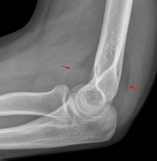
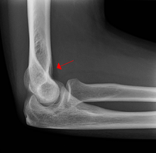
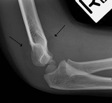
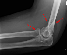
On an elbow X-ray, the fat pad sign, also known as the sail sign suggests an occult fracture. Its name derives from the fact that, it has the shape of a spinnaker (sail).[1] It is caused by displacement of the fat pad around the elbow joint. Both anterior and posterior fat pad signs exist, and both can be found on the same X-ray.
In children, a posterior fat pad sign suggests a condylar fracture of the humerus. In adults it suggests a radial head fracture. The fat pad sign only occurs after an intra-articular fracture.
The fat pad sign is invaluable in assessing for the presence of an intra-articular fracture of the elbow. An anterior fat pad is often normal. However a posterior fat pad seen on a lateral x-ray of the elbow is always abnormal. The patient will be unable to flex their elbow and requires orthopaedic input.[2]
Pathophysiology
The posterior fat pad is normally pressed in the olecranon fossa by the triceps tendon, and hence invisible on lateral radiograph of the elbow.[3] When there is a fracture of the distal head of the humerus, or other pathology involving the elbow joint, inflammation develops around the synovial membrane forcing the fat pad out of its normal physiologic resting place. This is visible as the "posterior fat pad sign" and is often the only visible marker of a fracture, particularly in the pediatrics population.
References
- ↑ Chapman S (1991). "The sail sign.". Br J Hosp Med 46 (6): 399–400. PMID 1760676.
- ↑ http://radiology.rsnajnls.org/cgi/content/full/222/2/419
- ↑ Goswami, MD, Gaurav K. "The Fat Pad Sign". Radiology. Retrieved 3 October 2012.