Eagle syndrome
| Eagle syndrome | |
|---|---|
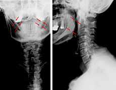
 Radiographs of the cervical spine: a-p and lateral view. Neither distinct malposition nor major degenerative changes of the cervical spine are recognizable. Formally and structurally inconspicuous cervical vertebral bodies and adnexa. But detection of a largely ossification of the ligamenta stylohyoidea on both sides. The patient's medical condition might be ascribed to a kerato-stylohyoidal syndrome. | |
| Classification and external resources | |
| DiseasesDB | 33542 |
| eMedicine | article/1447247 |
Eagle syndrome or styloid–carotid artery syndrome[1] is a rare condition where an elongated temporal styloid process (more than 30mm) is in conflict with the adjacent anatomical structures.
Two forms of eagle syndrome exists: The classic form and the vascular one.
Symptoms
Patients with this syndrome tend to be between 30 and 50 years of age but it has been recorded in teenagers and in patients > 75 years old. It is more common in women, with a male:female ratio ~ 1:2.
Patients with the classic "Eagle Syndrome" can present with unilateral sore throat, dysphagia, tinnitus, unilateral facial and neck pain, and otalgia.
In patients with the vascular form of "Eagle syndrome", the elongated styloid process is in contact with the extracranial internal carotid artery. This can cause a compression (while turning the head) or a dissection of the carotid artery causing a transient ischemic event or a stroke.
Diagnosis

Diagnosis is suspected when a patient presents with the symptoms of the classic form of "eagle syndrome" e.g. unilateral neck pain, sore throat or tinnitus. On the exam, one can sometimes palpate the tip of the styloid process in the back of the throat. The diagnosis of the vascular type is more difficult and requires an expert opinion. One should have a high level of suspicion when neurological symptoms occur upon head rotation. Symptoms tend to be worsened on bimanual palpation of the styloid through the tonsillar bed. They may be relieved by infiltration of lidocaine into the tonsillar bed. Because of the proximity of several large vascular structures in this area this procedure should not be considered to be risk free.
Imaging is important and is diagnostic. Visualizing the styloid process on a CT scan with 3D reconstruction is the suggested imaging technique.[2] The enlarged styloid may be visible on an orthopantogram or a lateral soft tissue X ray of the neck.
It is worth noting that the styloid may be enlarged (>30 millimeters in length) in 4% of the population and only a small minority (~4%) of people with enlarged styloids have symptoms.
Treatment
In both the classic and vascular form, the treatment is surgical.[3] A partial styloidectomy is the preferred approach. Repair of a damaged carotid artery is essential in order prevent further neurological complications.
History
The condition was first described by the American otorhinolaryngologist Watt Weems Eagle in 1937.[4]
Aetiology
Elongation of the styloid process and calcification of the stylohyoid ligament may be seen in patients who suffer from bruxism. There may also be desaturation of oxygen during sleep which is suggestive of sleep apnea.
References
- ↑ Hoffmann, E.; Räder, C.; Fuhrmann, H.; Maurer, P. (2013). "Styloid–carotid artery syndrome treated surgically with Piezosurgery: A case report and literature review". Journal of Cranio-Maxillofacial Surgery 41 (2): 162–166. doi:10.1016/j.jcms.2012.07.004. PMID 22902881.
- ↑ Karam C, Koussa S (December 2007). "[Eagle syndrome: the role of CT scan with 3D reconstructions]". J Neuroradiol (in French) 34 (5): 344–5. doi:10.1016/j.neurad.2007.08.001. PMID 17997158.
- ↑ Orhan KS, Güldiken Y, Ural HI, Cakmak A (April 2005). "[Elongated styloid process (Eagle's syndrome): literature review and a case report]". Agri (in Turkish) 17 (2): 23–5. PMID 15977090.
- ↑ Eagle's syndrome (Watt Weems Eagle) at Who Named It?
| Wikimedia Commons has media related to Eagle syndrome. |