Denticulate ligaments
| Denticulate ligaments | |
|---|---|
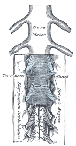 The medulla spinalis and its membranes. (Ligamentum denticulatum labeled vertically at bottom left.) | |
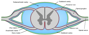 Diagrammatic transverse section of the medulla spinalis and its membranes. (Denticulate ligament not labeled, but region is visible.) | |
| Details | |
| Latin | ligamentum denticulatum |
| Identifiers | |
| Gray's | p.750 |
| Dorlands /Elsevier | l_09/12492205 |
| TA | A14.1.01.310 |
| FMA | 71245 |
| Anatomical terminology | |
The pia mater of the spinal cord has a pair of denticulate ligaments (one on each side of the spinal cord) with 21 attachments per side which attach it to the arachnoid and dura mater. Named for their tooth-like appearance, the denticulate ligaments are traditionally believed to provide stability for the spinal cord against motion within the vertebral column. Fortunately, from a clinical standpoint, denticulate ligaments do not play a significant role in lumbar spinal stenosis when compared to issues such as disc herniations, facet hypertrophy, shape of spinal canal, size of spinal canal, ligamentum flavum hypertrophy, or degenerative joint disease resulting in bony osteophyte formation. There are schools of thought that the anterior and posterior nerve roots are affected by abnormal tensions in the denticulate ligaments.[1]
Structure
Each denticulate ligament is composed of a single narrow fibrous strip that extends from the craniovertebral junction to T12. Each ligament features 18-20 triangular extensions that attach to the dura at their apices. The triangular extensions are smaller and more numerous at the cervical levels, and are larger and less numerous at the thoracic levels. The apices of the extensions attach to the dura via fibrous bands at cervical levels (each band 3–5 mm (0.12–0.20 in) long) and lower thoracic levels (21–26 mm (0.83–1.02 in) long), whereas they attach directly to the dura at upper thoracic levels.
The narrow fibrous strip of the denticulate ligament features longitudinally oriented collagen fibers, whereas the triangular extensions are composed of transverse and obliquely oriented collagen fibers. The collagen fibers are thicker and more abundant at the cervical than at the thoracic levels.
These ligaments may be affected by altered motion and position of the vertebral segments
Additional images
-

A portion of the spinal cord, showing its right lateral surface. The dura is opened and arranged to show the nerve roots.
Sources
- Tubbs RS, Salter G, Grabb PA, Oakes WJ (April 2001). "The denticulate ligament: anatomy and functional significance". Journal of Neurosurgery 94 (2 Suppl): 271–5. doi:10.3171/spi.2001.94.2.0271. PMID 11302630.
- Clinically Oriented Anatomy. Moore, Keith and Arthur F. Dalley. Philadelphia, Lipincott, Williams and Williams 2006.
- RC Schafer DC PhD. Basic Principles of Chiropractic Neuroscience - Chapter 5; Neuroconceptual Models of Chiropractic ACA Press 1998 | http://www.chiro.org/ACAPress/Neuroconceptual_Models_of_Chiropractic.html''.
- Ceylan D, Tatarlı N, Abdullaev T et al. (July 2012). "The denticulate ligament: anatomical properties, functional and clinical significance". Acta Neurochirurgica 154 (7): 1229–34. doi:10.1007/s00701-012-1361-x. PMID 22555553.
See also
- This article uses anatomical terminology; for an overview, see anatomical terminology.
References
- ↑ Schafer. "NEUROCONCEPTUAL MODELS OF CHIROPRACTIC". Retrieved 11 February 2014.
External links
- denticulate+ligament at eMedicine Dictionary
- Anatomy figure: 02:05-03 at Human Anatomy Online, SUNY Downstate Medical Center - "Coverings of the spinal cord."
| ||||||||||||||||||||||||||||||||||||||||||||||||