Cypovirus
| Cypovirus | |
|---|---|
 | |
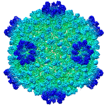
| Structure of a transcribing cypovirus. EMDB entry EMD-5376.[1] | |
| Virus classification | |
| Group: | Group III (dsRNA) |
| Order: | Unassigned |
| Family: | Reoviridae |
| Subfamily: | Spinareovirinae |
| Genus: | Cypovirus |
| Type species | |
| Cypovirus 1 | |
| Species | |
| |
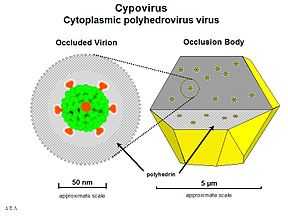
Cypovirus (CPV), short for cytoplasmic polyhedrosis virus, is a genus of viruses in the Reoviridae family. The virions have an icosahedral structure typical of other reoviruses and are 55–69 nm in diameter. The genome is composed of 10 segments of double-stranded RNA. The virions are embedded in a protein matrix to form the structures referred to as polyhedra or occlusion bodies.
Cypoviruses have only been isolated from insects. Morphologically, these viruses have much in common with the much more widely studied nucleopolyhedroviruses (NPV), a genus of arthropod viruses in the Baculovirus family. However, CPV have an RNA genome and replicate in the cytoplasm of the infected cells while NPV have a DNA genome and replicate in the nucleus.
Structure and proteins

CPVs are classified into 14 species based on the electrophoretic migration profiles of their genome segments. Cypovirus has only a single capsid shell, which is similar to the orthoreovirus inner core. CPV exhibits striking capsid stability and is fully capable of endogenous RNA transcription and processing. The overall folds of CPV proteins are similar to those of other reoviruses. However, CPV proteins have insertional domains and unique structures that contribute to their extensive intermolecular interactions. The CPV turret protein contains two methylase domains with a highly conserved helix-pair/β-sheet/helix-pair sandwich fold but lacks the β-barrel flap present in orthoreovirus λ2. The stacking of turret protein functional domains and the presence of constrictions and A spikes along the mRNA release pathway indicate a mechanism that uses pores and channels to regulate the highly coordinated steps of RNA transcription, processing, and release.[2]
Pathogenesis
Infection occurs when a susceptible insect consumes the polyhedra, usually as a contaminant on the insect’s food (in most cases, foliage of a plant). The polyhedra dissolve in the digestive tract of the insect, releasing the virus particles that penetrate the gut epithelial cells. Replication of the virus is often confined to these cells and the progeny virus, in the form of new polyhedra are excreted in the insect feces, thus contaminating more foliage resulting in the spread of the disease to additional insects. The progression of the disease can be rather slow, but the virus infection is normally fatal.
See also
References
- ↑ Yang, C.; Ji, G.; Liu, H.; Zhang, K.; Liu, G.; Sun, F.; Zhu, P.; Cheng, L. (2012). "Cryo-EM structure of a transcribing cypovirus". Proceedings of the National Academy of Sciences 109 (16): 6118. doi:10.1073/pnas.1200206109.
- ↑ Zhou ZH (2008). "Cypovirus". Segmented Double-stranded RNA Viruses: Structure and Molecular Biology. Caister Academic Press. ISBN 978-1-904455-21-9.