Coccygeal glomus
| Coccygeal glomus | |
|---|---|
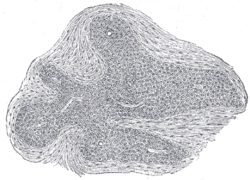
 Section of an irregular nodule of the glomus coccygeum. X 85. The section shows the fibrous covering of the nodule, the bloodvessels within it, and the epithelial cells of which it is constituted. | |
| Details | |
| Latin | glomus coccygeum |
| median sacral artery | |
| Identifiers | |
| Gray's | p.1281 |
| Dorlands /Elsevier | g_07/12394809 |
| TA | A12.2.12.011 |
| FMA | 15649 |
| Anatomical terminology | |
The coccygeal glomus (coccygeal gland or body; Luschka’s gland) is a vestigial structure[1] placed in front of, or immediately below, the tip of the coccyx.
Anatomy
It is about 2.5 mm. in diameter and is irregularly oval in shape; several smaller nodules are found around or near the main mass.
It consists of irregular masses of round or polyhedral cells, the cells of each mass being grouped around a dilated sinusoidal capillary vessel.
Each cell contains a large round or oval nucleus, the protoplasm surrounding which is clear, and is not stained by chromic salts.
Clinical significance
It may appear similar to a glomus tumor.[2]
References
This article incorporates text in the public domain from the 20th edition of Gray's Anatomy (1918)
- ↑ Rahemtullah A., Szyfelbein K, Zembowicz A. (2005). "Glomus coccygeum: report of a case and review of the literature.". Retrieved 21 October 2014.
- ↑ Santos L, Chow C, Kennerson A (2002). "Glomus coccygeum may mimic glomus tumour.". Pathology 34 (4): 339–43. doi:10.1080/003130202760120508. PMID 12190292.
| ||||||||||||||||||||||||||||||||||||||||||||||||||||||||||||||||||||||||||||||||||||||||||||