Clavicle fracture
| Clavicle Fracture | |
|---|---|
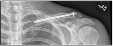
 X-ray of a left clavicle fracture | |
| Classification and external resources | |
| ICD-10 | S42.0 |
| ICD-9 | 810 |
| MedlinePlus | 001588 |
| eMedicine | orthoped/50 |
A clavicle fracture is a bone fracture in the clavicle, or collarbone. It is often caused by a fall onto an outstretched upper extremity, a fall onto a shoulder, or a direct blow to the clavicle. Many research projects are underway regarding the medical healing process of clavicle fractures.
Anatomy
The clavicle is the bone that connects the trunk of the body to the arm, and it is located directly above the first rib. There is a clavicle on each side of the front, upper part of the chest. The clavicle consists of a medial end, shaft, and a lateral end. The medial end connects with the manubrium of the sternum and gives attachments to the fibrous capsule of the sternoclavicular joint, articular disc, and interclavicular ligament. The lateral end connects at the acromion of the scapula which is referred to as the acromioclavicular joint. The clavicle forms a slight S-shaped curve where it curves from the sternal end laterally and anteriorly for near half its length, then forming a posterior curve to the acromion of the scapula.

Risk factors/prevention
Those who have a low dietary intake of calcium and Vitamin D may have a higher risk of clavicular fractures. Increasing the integrity of the bone by a sufficient amount of dietary calcium and vitamin D will help to prevent fractures in the bone. Also, sedentary individuals may be at a higher risk due to weakness in muscle stabilizers of the clavicle. Also, participation in extreme sports such as mountain biking and snowboarding will increase the risk of a clavicular fracture as well.
Mechanism of injury
Clavicle fractures are commonly known as a breaking of the collarbone, and they are usually a result of injury or trauma. The most common type of fractures occur when a person falls horizontally on the shoulder or with an outstretched hand. A direct hit to the collarbone will also cause a break. In most cases, the direct hit occurs from the lateral side towards the medial side of the bone. The muscles involved in clavicle fractures include the deltoid, trapezius, subclavius, sternocleidomastoid, sternohyoid and pectoralis major muscles. The ligaments involved include the Conoid ligament and Trapezoid ligament. Incidents that may lead to a clavicle fracture include automobile accidents, biking accidents, especially common in mountain biking, horizontal falls on the shoulder joint, or contact sports such as football,rugby or wrestling.
Signs and symptoms
- Pain, particularly with upper extremity movement or on front part of upper chest.
- Swelling
- Often, after the swelling has subsided, the fracture can be felt through the skin.
- Sharp pain when any movement is made.
- Referred pain: dull to extreme ache in and around clavicle area, including surrounding muscles.
- Possible nausea, dizziness, and/or spotty vision due to extreme pain
Diagnosis
The basic method to check for a clavicle fracture is by an X-Ray of the clavicle in order to determine the fracture type and extent of injury. In most cases, x-rays will be taken of both clavicle bones for comparison purposes. In more severe cases a computerized tomography scan (CAT scan) or Magnetic resonance imaging scan (MRI) will be taken. However, the standard method of diagnosis is through ultrasound imaging performed in the emergency room may be equally accurate in children.[1]
Treatment
Medication can be prescribed to ease the pain as well as antibiotic or tetanus shot for any skin breaks. In severe cases, surgery may be needed in order to place bones in normal positions utilizing pins, plates, and screws to hold bones together.
Nonoperative
The arm must be supported by use of a splint or sling to keep the joint stable and decrease the risk of further damage. Usually, a figure-of-eight splint that wraps the shoulders to keep them forced back is used and the arm is placed in a clavicle strap for comfort.
Current practice is generally to provide a sling, and pain relief, and to allow the bone to heal itself, monitoring progress with X-rays every week or few weeks. Surgery is employed in 5-10% of cases. However, a recent study supports primary plate fixation of completely displaced midshaft clavicular fractures in active adult patients.[2]
If the fracture is at the lateral end, the risk of nonunion is greater than if the fracture was of the shaft.[3]
Surgical
The surgery is indicated when one or more of the following conditions presents.
- Comminution with separation (multiple piece)
- Significant Foreshortening of the clavicle (indicated by shoulder forward).
- Skin penetration (Open Fracture).
- Clearly associated nervous and vascular trauma (Brachial Plexus or Supra Clavicular Nerves).
- Non Union after several months (3–6 months, typically)
- Distal Third Fractures ( high risk of non-union)
A discontinuity in the bone shape often results from a clavicular fracture, visible through the skin, if not treated with surgery. Surgical procedure will often call for ORIF (Open Reduction Internal [plate] Fixation) where an anatomically shaped titanium or steel plate is affixed along the superior aspect of the bone via several screws. In some cases the plate may be removed after healing, but this is very rarely required (based on nerve interaction or tissue aggravation), and typically considered an elective procedure. Alternatively, intramedullary fixation devices (within the medullary canal) can be implanted to support the fracture during healing. These devices are implanted within the clavicle's canal to support the bone from the inside. Typical surgical complications are infection, neurological symptoms distal the incision (sometimes to the extremity), and non-union.
Prognosis
Healing time varies based on age, health, complexity and location of the break as well as the bone displacement. For adults, a minimum of 2–6 weeks of sling immobilization is normally employed to allow initial bone and soft tissue healing, teenagers require slightly less, children can often achieve the same level in two weeks. During this period, patients may remove the sling to practice passive pendulum Range of Motion (ROM) exercises to reduce atrophy in the elbow and shoulder, but they are minimized to 15-20 degrees off vertical. Depending on the severity of fracture, a person can begin to use the arm if comfortable with movement and no pain results. The final goal is to be able to have full range of motion with no pain; therefore, if any pain exists, it is best to allow for more recovery time. Depending on severity of the fracture, athletes involved in contact sports may need a longer period of rest to heal to avoid re-fracturing bone. A person should be able to return unrestricted to any sports or work by 3 months after the injury.
Rehab Protocol
General considerations
- Do not elevate surgical arm above 70º in any plane for the first 4 weeks post-op
- Do not lift any objects over 5 pounds with the surgical arm for the first 6 weeks
- Avoid repeated reaching for the first 6 weeks
- Ice shoulder 3 - 5 times (15 minutes each time) per day to control swelling and inflammation
- An arm sling is used for 3 - 4 weeks post-op
- Maintain good upright shoulder girdle posture at all times and especially during sling use
- Intermittent X-ray to monitor healing as needed
- MD follow-up visits at Day 1, Day 14 with nurse for suture removal, Month 1, Month 3, and 1 Year Post-op
Week 1
- MD visit Day 1 post-op to change dressing and review home program
- Soft tissue treatments for associated shoulder and neck musculature for comfort
- Cardiovascular training such as stationary bike throughout rehabilitation period
- Exercises (3 times per day):
- Pendulum exercises
- Squeeze ball
- Triceps with Theraband
- Isometric rotator cuff external and internal rotations with arm at side
- Isometric shoulder abduction, adduction, extension, and flexion with arm at side
Weeks 2 – 4
- Soft tissue treatments for associated shoulder and neck musculature for comfort
- Gentle pulley for shoulder ROM 2 times/day
- Elbow pivots PNF, wrist PNF
- Isometric scapular PNF, mid-range
Weeks 4 – 8
- MD visit and will usually be progressed to a more aggressive ROM and strength program
- At Week 4:
- Start mid-range of motion (ROM) rotator cuff external and internal rotations and active and light resistance exercises (through 75% of ROM as patient's symptoms permit) without shoulder elevation and avoiding extreme end ROM
- Strive for progressive gains to active 90º of shoulder flexion and abduction
Weeks 8 – 12
- Seek full shoulder active ROM in all planes
- Increase manual mobilizations of soft tissue as well as glenohumeral and scapulothoracic joints for ROM
- No repeated heavy resisted exercises or lifting until 3 months
Week 12 post-op and beyond
- Start a more aggressive strengthening program as tolerated
Increase the intensity of strength and functional training for gradual return to activities and sports
- Return to specific sports is determined by the physical therapist through functional testing specific to the injury
Epidemiology
Clavicle fractures occur 30-64 cases per 100,000 a year and are responsible for 2.6-5% of all fractures.[4] This type of fracture occurs more often in males.[4] About half of all clavicle fractures occur in children under the age of 7 and is the most common pediatric fracture. Clavicle fractures involve approximately 5% of all fractures seen in hospital emergency admissions. Clavicles are the most common broken bone in the human body.[5] It is most often fractured in the middle third of its length which is its weakest point. The lateral fragment is depressed by the weight of the arm and is pulled medially and forward by the strong adductor muscles of the shoulder joint, especially the pectoralis major. The part of the clavicle near the center of the body is tilted upwards by the sternocleidomastoid muscle. Children and infants are particularly prone to it. Newborns often present clavicle fractures following a difficult delivery.
After fracture of the clavicle, the sternocleidomastoid muscle elevates the medial fragment of the bone. The trapezius muscle is unable to hold up the distal fragment owing to the weight of the upper limb, and thus the shoulder droops. The adductor muscles of the arm, such as the pectoralis major, may pull the distal fragment medially causing the bone fragments to override.
History
Hippocrates, 4th century BC:
When, then, a [clavicle] fracture has recently taken place, the patients attach much importance to it, as supposing the mischief greater than it really is, and the physicians bestow great pains in order that it may be properly bandaged; but in a little time the patients, having no pain, nor finding any impediment to their walking or eating, become negligent; and the physicians finding they cannot make the parts look well, take themselves off, and are not sorry at the neglect of the patient, and in the meantime the callus is quickly formed.'
The management of skeletal injuries in ancient Egypt – Collar bone:
If thou examinest a man having a break in his collar bone and shouldst thou find his collar bone short and separated from its fellow, I will treat. Place him prostrate on his back with something folded between his shoulder blades; thou shouldst spread out with his two shoulders to stretch apart his collar bone until the break falls in its place.
See also
References
- ↑ Cross KP, Warkentine FH, Kim IK, Gracely E, Paul RI (July 2010). "Bedside ultrasound diagnosis of clavicle fractures in the pediatric emergency department". Acad Emerg Med 17 (7): 687–93. doi:10.1111/j.1553-2712.2010.00788.x. PMID 20653581.
- ↑ Nonoperative Treatment Compared with Plate Fixation of Displaced Midshaft Clavicular Fractures - http://www.ejbjs.org/cgi/content/abstract/89/1/1
- ↑ Khan LA, Bradnock TJ, Scott C, Robinson CM (February 2009). "Fractures of the clavicle". J Bone Joint Surg Am 91 (2): 447–60. doi:10.2106/JBJS.H.00034. PMID 19181992.
- ↑ 4.0 4.1 Malik S, Chiampas G, Leonard H (November 2010). "Emergent evaluation of injuries to the shoulder, clavicle, and humerus". Emerg Med Clin North Am 28 (4): 739–63. doi:10.1016/j.emc.2010.06.006. PMID 20971390.
- ↑ Richard S. Snell, MD, PHD (2010-03-10). "Chapter 9: The upper Limb". Clinical Anatomy by Regions (8th ed.). Lippincott Williams & Wilkins. p. 433. ISBN 978-0-7817-6404-9.
| ||||||||||||||||||||||||||||||||||||||||||||||||||||||||||||||||||||||||||||||||||||||||||||||||||