Cauda equina
| Cauda equina | |
|---|---|
|
Cauda equina and filum terminale seen from behind. | |
|
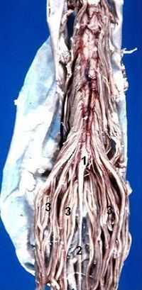
Human caudal spinal cord anterior view | |
| Details | |
| Latin | Cauda equina |
| Iliolumbar artery | |
| Identifiers | |
| Gray's | p.919 |
| MeSH | A08.800.800.720.725.150 |
| TA | A14.2.00.036 |
| FMA | 52590 |
| Anatomical terminology | |
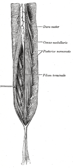
The cauda equina (Latin for "horse's tail") is a bundle of spinal nerves and spinal nerve roots, consisting of the second through fifth lumbar nerve pairs, the first through fifth sacral nerve pairs, and the coccygeal nerve, all of which originate in the conus medullaris of the spinal cord. The nerves that compose the cauda equina innervate the pelvic organs and lower limbs to include motor innervation of the hips, knees, ankles, feet, internal anal sphincter and external anal sphincter. In addition, the cauda equina extends to sensory innervation of the perineum and, partially, parasympathetic innervation of the bladder.[1]
Structure
Development
In humans, the spinal cord stops growing in infancy and the end of the spinal cord is about the level of the third lumbar vertebra, or L3, at birth. By the time adulthood is reached, because the bones of the vertebral column continue to grow, the end of the cord is at the level of L1 or L2 (closer to the head). However due to normal anatomical variation, the cord may end anywhere between L3 and the twelfth thoracic vertebra (T12) in adults. Individual spinal nerve roots arise from the cord as they do closer to the head, but as the differential growth occurs, the top end of the nerve stays attached to the spinal cord while the lower end of the nerve exits the spinal column at its proper level. This results in a "bundle"-like structure of nerve fibers that extends caudally from the end of the spinal cord, gradually declining in number further down as individual pairs leave the spinal column.
Clinical significance
The cauda equina exists within the lumbar cistern, which is the space formed from the surrounding dural sac. Cerebrospinal fluid is drawn from this space during a lumbar puncture.
History
The cauda equina was named after its resemblance to a horse's tail (Latin: cauda equina) by the French anatomist Andreas Lazarius (André du Laurens) in the 17th century.
See also
- This article uses anatomical terminology; for an overview, see anatomical terminology.
References
Saladin, Kenneth S. Anatomy and Physiology The Unity of Form and Function
External links
- VIRTUAL Spine - Online Learning Resource
- MedlinePlus Image 19504
- Anatomy photo:02:08-0106 at the SUNY Downstate Medical Center
- Cross section image: pembody/body12a - Plastination Laboratory at the Medical University of Vienna
- Dissection of deep back and spinal cord video: great view of the Cauda Equina
| ||||||||||||||||||||||||||||||||||||||||||||||||||||||||||||||||||||||||||||