Capitate bone
| Capitate bone | |
|---|---|
_01_palmar_view.png) Left hand anterior view (palmar view). Capitate-bone shown in red. | |
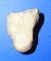
 The left capitate bone. Left: ulnar surface (little-finger-side surface). Right: radial surface (thumb-side surface) | |
| Details | |
| Latin | Os capitatum; os magnum |
| Identifiers | |
| Gray's | p.226 |
| MeSH | A02.835.232.087.319.150.150 |
| Dorlands /Elsevier | o_07/12598140 |
| TA | A02.4.08.011 |
| FMA | 23727 |
| Anatomical terms of bone | |
The capitate bone /ˈkæpɨteɪt/ is the largest of the carpal bones in the human hand, and occupies the center of the wrist. It presents, above, a rounded portion or head, which is received into the concavity formed by the scaphoid and lunate bones; a constricted portion or neck; and below this, the body.[1] The bone is also found in many other mammals, and is homologous with the "third distal carpal" of reptiles and amphibians.
Structure
The capitate is the largest carpal bone found within the hand.[2] The capitate is found within the distal row of carpal bones. It is the largest carpal bone. The capitate lies directly adjacent to the metacarpal of the ring finger on its distal surface, has the hamate on its ulnar surface and trapezoid on its radial surface, and abuts the lunate and scaphoid proximally.[3] :708–709
Surfaces
The superior surface is round, smooth, and articulates with the lunate bone.[1]
The inferior surface is divided by two ridges into three facets, for articulation with the second, third, and fourth metacarpal bones, that for the third being the largest.[1]
The dorsal surface is broad and rough.[1]
The palmar surface is narrow, rounded, and rough, for the attachment of ligaments and a part of the adductor pollicis muscle.[1]
The lateral surface articulates with the lesser multangular by a small facet at its anterior inferior angle, behind which is a rough depression for the attachment of an interosseous ligament. Above this is a deep, rough groove, forming part of the neck, and serving for the attachment of ligaments; it is bounded superiorly by a smooth, convex surface, for articulation with the scaphoid bone.[1]
The medial surface articulates with the hamate bone by a smooth, concave, oblong facet, which occupies its posterior and superior parts; it is rough in front, for the attachment of an interosseous ligament.[1]
Variation
The capitate bone variably articulates with the metacarpal of the index finger.[2]
Ossification
The ossification of capitate starts at 1 - 5 months.[4]
Function
The carpal bones function as a unit to provide a bony superstructure for the hand.[3] :708
History
The etymology derives from the Latin Latin: capitātus, "having a head," from Latin: capit-, meaning "head."[5]
Additional images
-
_-_animation01.gif)
Position of capitate bone (shown in red). Left hand. Animation.
-
_-_animation02.gif)
Capitate bone of the left hand. Close up. Animation.
-

Capitate bone of the left hand. Ulnar surface (little-finger-side surface)
-

Capitate bone of the left hand. Radial surface (thumb-side surface)
-

Right hand posterior view (dorsal view). Thumb on bottom.
-

Right hand anterior view (palmar view). Thumb on top.
-

Capitate bone shown in yellow. Left hand. Palmar surface.
-

Capitate bone shown in yellow. Left hand. Dorsal surface.
-

Transverse section across the wrist (palm on top, thumb on left). Capitate bone shown in yellow.
-

Cross section of wrist (thumb on left). Capitate shown in red.
See also
| Wikimedia Commons has media related to Capitate bone. |
- This article uses anatomical terminology; for an overview, see anatomical terminology.
- Carpal bone
References
This article incorporates text in the public domain from the 20th edition of Gray's Anatomy (1918)
- ↑ 1.0 1.1 1.2 1.3 1.4 1.5 1.6 Gray's Anatomy (1918). See infobox.
- ↑ 2.0 2.1 Eathorne, SW (Mar 2005). "The wrist: clinical anatomy and physical examination--an update.". Primary care 32 (1): 17–33. doi:10.1016/j.pop.2004.11.009. PMID 15831311.
- ↑ 3.0 3.1 Drake, Richard L.; Vogl, Wayne; Tibbitts, Adam W.M. Mitchell; illustrations by Richard; Richardson, Paul (2005). Gray's anatomy for students. Philadelphia: Elsevier/Churchill Livingstone. ISBN 978-0-8089-2306-0.
- ↑ Balachandran, Ajay; Kartha, Moumitha; Krishna, Anooj; Thomas, Jerry; K, Prathilash; TN, Prem; GK, Libu; B, Krishnan; John, Liza (2014). "A Study of Ossification of Capitate, Hamate, Triquetral & Lunate in Forensic Age Estimation". Indian Journal of Forensic Medicine & Toxicology 8 (2): 218–224. doi:10.5958/0973-9130.2014.00720.8. ISSN 0973-9130. Retrieved 18 August 2014.
- ↑ Harper, Douglas. "Capitate". Online Etymology Dictionary. Retrieved 5 January 2014.
| ||||||||||||||||||||||||||||||||||||||||||||||||||||||||