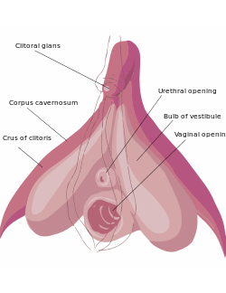
Bulb of vestibule
| Vestibular bulbs | |
|---|---|
 The internal anatomy of the human vulva, with the clitoral hood and labia minora indicated as lines | |
 | |
| Details | |
| Latin | bulbus vestibuli vaginae |
| artery of bulb of vestibule | |
| vein of bulb of vestibule | |
| superficial inguinal lymph nodes | |
| Identifiers | |
| Gray's | p.1266 |
| Dorlands /Elsevier | 12200326 |
| TA | A09.2.01.013 |
| FMA | 20199 |
| Anatomical terminology | |

In female anatomy, the vestibular bulbs, also known as the clitoral bulbs, are aggregations of erectile tissue that are an internal part of the clitoris. They can also be found throughout the vestibule—next to the clitoral body, clitoral crura, urethra, urethral sponge, and vagina.
They are to the left and right of the urethra, urethral sponge, and vagina.
The vestibular bulbs are homologous to the bulb of penis and adjoining part of the corpus spongiosum of the male, and consists of two elongated masses of erectile tissue, placed one on either side of the vaginal orifice and united to each other in front by a narrow median band termed the pars intermedia.
Their posterior ends are expanded and are in contact with the greater vestibular glands; their anterior ends are tapered and joined to one another by the pars intermedia; their deep surfaces are in contact with the inferior fascia of the urogenital diaphragm; superficially they are covered by the bulbospongiosus.
Physiology
During the response to sexual arousal the bulbs fill with blood, which then becomes trapped, causing erection. As the clitoral bulbs fill with blood, they tightly cuff the vaginal opening, causing the vulva to expand outward. Nearby structures this may put pressure on include the corpus cavernosum of clitoris and crus of clitoris.
The blood inside the bulb’s erectile tissue is released to the circulatory system by the spasms of orgasm, but if orgasm does not occur, the blood will exit the bulbs over several hours.[1]
References
This article incorporates text in the public domain from the 20th edition of Gray's Anatomy (1918)
External links
- Anatomy photo:41:11-0204 at the SUNY Downstate Medical Center - "The Female Perineum: Muscles of the Superficial Perineal Pouch"
- figures/chapter_38/38-4.HTM — Basic Human Anatomy at Dartmouth Medical School
| ||||||||||||||||||||||||||||||||||||||||||||||||||||||||||||||||||||||||||||||||||||||||