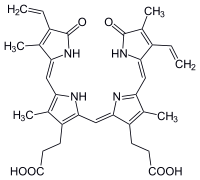
Biliverdin
 | |
| Identifiers | |
|---|---|
| 114-25-0 | |
| ChEBI | CHEBI:17033 |
| ChEMBL | ChEMBL455477 |
| ChemSpider | 10628548 |
| |
| Jmol-3D images | Image |
| MeSH | Biliverdin |
| PubChem | 251 |
| |
| Properties | |
| C33H34N4O6 | |
| Molar mass | 582.646 |
| Melting point | > 300 °C |
| Hazards | |
| MSDS | Sigma-Aldrich |
| Main hazards | Irritant |
| Except where noted otherwise, data is given for materials in their standard state (at 25 °C (77 °F), 100 kPa) | |
| | |
| Infobox references | |
Biliverdin is a green tetrapyrrolic bile pigment, and is a product of heme catabolism.[1][2] It is the pigment responsible for a greenish color sometimes seen in bruises.[2]
Metabolism

Biliverdin results from the breakdown of the heme moiety of hemoglobin in erythrocytes. Macrophages break down senescent erythrocytes and break the heme down into biliverdin, which normally rapidly reduces to free bilirubin.[1][3] Biliverdin is seen briefly in some bruises as a green color. In bruises, its breakdown into bilirubin leads to a yellowish color.[2]
Role in disease
Biliverdin has been found in excess in the blood of humans suffering from hepatic diseases. Jaundice is caused by the accumulation of biliverdin or bilirubin (or both) in the circulatory system and tissues.[1] Jaundiced skin and sclera (whites of the eyes) are characteristic of liver failure.
Role in treatment of disease
While typically regarded as a mere waste product of heme breakdown, evidence that suggests that biliverdin — and other bile pigments — has a physiological role in humans has been mounting.[4][5]
Bile pigments such as biliverdin possess significant anti-mutagenic and antioxidant properties and therefore may fulfil a useful physiological function.[5] Biliverdin and bilirubin have been shown to be potent scavengers of peroxyl radicals.[4][5] They have also been shown to inhibit the effects of polycyclic aromatic hydrocarbons, heterocyclic amines, and oxidants — all of which are mutagens. Some studies have found that people with higher concentration levels of bilirubin and biliverdin in their bodies have a lower frequency of cancer and cardiovascular disease.[4] It has been suggested that biliverdin — as well as many other tetrapyrrolic pigments — may function as an HIV-1 protease inhibitor[6] as well as having beneficial effects in asthma[5] though further research is needed to confirm these results. There are currently no practical implications for using biliverdin in the treatment of any disease.
In non-human animals
Biliverdin is an important component of avian egg shells. There is a significantly higher concentration of biliverdin in blue egg shells than in brown egg shells. Research has shown that the biliverdin of egg shells is produced from the shell gland, rather than from the breakdown of erythrocytes in the blood stream. The presence of biliverdin in egg shells may be an indicator of female fitness, and therefore likely demonstrates evolutionary significance.[7]
Along with its presence in avian egg shells, other studies have also shown that biliverdin is present in the blue-green blood of many marine fish, the blood of tobacco hornworm, the wings of moth and butterfly, the serum and eggs of frogs, and the placenta of dogs.[8] In the garfish (Belone belone) and related species, the bones are bright green because of biliverdin.
Biliverdin is also present in the green blood, muscles, bones, and mucosal lining of skinks of the genus Prasinohaema, found in New Guinea. It is uncertain whether this presence of biliverdin is an ecological or physiological adaptation of any kind. It has been suggested that accumulation of biliverdin might deter harmful infection by Plasmodium malaria parasites, though no statistically significant correlation has been established.[9] The Cambodian frog, Chiromantis samkosensis also exhibits this character along with turquoise bones.[10]
In fluorescence imaging
In a complex with reengineered bacterial phytochrome, biliverdin has been employed as an IR-emitting chromophore for in vivo imaging.[11][12] In contrast to fluorescent proteins which form their chromophore through posttranslational modifications of the polypeptide chain, phytochromes bind an external ligand (in this case, biliverdin), and successful imaging of the first bacteriophytochrome-based probe required addition of the exogenous biliverdin.[11] Recent studies demonstrated that bacteriophytochrome-based fluorescent proteins with high affinity to biliverdin can be imaged in vivo utilizing endogenous ligand only and, thus, with the same ease as the conventional fluorescent proteins.[12] Advent of the second and further generations of the biliverdin-binding bacteriophytochrome-based probes should broaden the possibilities for the non-invasive in vivo imaging.
See also
References
- ↑ 1.0 1.1 1.2 Boron W, Boulpaep E. Medical Physiology: a cellular and molecular approach, 2005. 984-986. Elsevier Saunders, United States. ISBN 1-4160-2328-3
- ↑ 2.0 2.1 2.2 Mosqueda L, Burnight K, Liao S (2005). "The Life Cycle of Bruises in Older Adults". Journal of the American Geriatrics Society. 53(8):1339-1343. doi:10.1111/j.1532-5415.2005.53406.x
- ↑ Seyfried, H; Klicpera, M; Leithner, C; Penner, E (1976). "Bilirubin metabolism (author's transl)". Wiener klinische Wochenschrift 88 (15): 477–82. PMID 793184.
- ↑ 4.0 4.1 4.2 Bulmer, AC; Ried, K; Blanchfield, JT; Wagner, KH (2008). "The anti-mutagenic properties of bile pigments". Mutation research 658 (1–2): 28–41. doi:10.1016/j.mrrev.2007.05.001. PMID 17602853.
- ↑ 5.0 5.1 5.2 5.3 Ohrui, T; Yasuda, H; Yamaya, M; Matsui, T; Sasaki, H (2003). "Transient relief of asthma symptoms during jaundice: a possible beneficial role of bilirubin". The Tohoku journal of experimental medicine 199 (3): 193–6. doi:10.1620/tjem.199.193. PMID 12703664.
- ↑ McPhee, F; Caldera, PS; Bemis, GW; McDonagh, AF; Kuntz, ID; Craik, CS (1996). "Bile pigments as HIV-1 protease inhibitors and their effects on HIV-1 viral maturation and infectivity in vitro". The Biochemical journal. 320 ( Pt 2) (Pt 2): 681–6. PMC 1217983. PMID 8973584.
- ↑ Lote, CJ; Saunders, H (1991). "Aluminium: gastrointestinal absorption and renal excretion". Clinical science (London, England : 1979) 81 (3): 289–95. PMID 1655328.
- ↑ Fang, LS; Bada, JL (1990). "The blue-green blood plasma of marine fish". Comparative biochemistry and physiology. B, Comparative biochemistry 97 (1): 37–45. doi:10.1016/0305-0491(90)90174-R. PMID 2253479.
- ↑ Austin C, Perkins S (2006). "Parasites in a biodiversity hotspot: a survey of hematozoa and a molecular phyolgenetic analysis of plasmodium in New Guinea skinks". Journal of Parasitology 92(4):770-777. doi:10.1645/GE-693R.1
- ↑ Lee Grismer, L.; Thy, Neang; Chav, Thou; Holden, Jeremy (2007). "A New Species of Chiromantis Peters 1854 (Anura: Rhacophoridae) from Phnom Samkos in the Northwestern Cardamom Mountains, Cambodia". Herpetologica 63 (3): 392. doi:10.1655/0018-0831(2007)63[392:ANSOCP]2.0.CO;2.
- ↑ 11.0 11.1 X. Shu et al. (2009). "Mammalian expression of infrared fluorescent proteins engineered from a bacterial phytochrome". Science 324 (5928): 804–807. doi:10.1126/science.1168683. PMC 2763207. PMID 19423828.
- ↑ 12.0 12.1 G.S.Filonov; Piatkevich, Kiryl D; Ting, Li-Min; Zhang, Jinghang; Kim, Kami; Verkhusha, Vladislav V et al. (2011). "Bright and stable near infra-red fluorescent protein for in vivo imaging". Nat Biotechnol 29 (8): 757–761. doi:10.1038/nbt.1918. PMC 3152693. PMID 21765402.
External links
| ||||||||||||||||||||||||||||||||||||||||||||||||||||||||||
| ||||||||||||||||||||||||||||||||||||||||||||||||||||||||||||||||||||||||||||||||||