Beta adrenergic receptor kinase


Beta adrenergic receptor kinase (also referred to as βARK or BARK) is a serine/threonine intracellular kinase. It is activated by PKA and its target is the beta adrenergic receptor. It is one method by which the cell will desensitize itself from epinephrine overstimulation.[1][2]
βARK Activation
- (steps 1) Upon stimulation of the Beta adrenergic receptor by epinephrine, Gs will be activated.
- (steps 2 and 3) Gs alpha will then stimulate adenyl cyclase to make cAMP.
- (steps 3 through 6) cAMP will then activate cAMP-dependent kinase (PKA), which, among other proteins that it acts on, will phosphorylate serine and threonine residues on βARK.
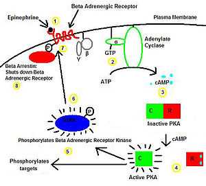
- (step 7) βARK, itself a serine/threonine kinase, will then phosphorylate serine and threonine resides on the β-adrenergic receptor itself.
- (step 8) This will facilitate Beta-arrestin's binding to the receptor. Additional stimulation by epinephrine will now be unable to activate Gs due to arrestin.
Therefore, βARK is a negative feedback enzyme that will prevent overstimulation of the β-adrenergic receptor.[1][2]
Other similar systems
In the rhodopsin system, which regulates rod cell function in the retina, rhodopsin kinase will phosphorylate serine and threonine residues on the rhodopsin receptor. Similarly to the βARK system, the phosphorylated rhodopsin residues will then bind to arrestin, resulting in receptor desensitization.
Structure
Protein Structure
The structure of βARK1 consists of a protein of 689 amino acids (79.7 kilodaltons) with a protein kinase catalytic domain that bears greatest sequence similarity to protein kinase C and the cyclic adenosine monophosphate (cyclic AMP)-dependent protein kinase.[3]
Gene structure
The gene spans approximately 23 kilobases and is composed of 21 exons interrupted by 20 introns. Exon sizes range from 52 bases (exon 7) to over 1200 bases (exon 21), intron sizes from 68 bases (intron L) to 10.8 kilobases (intron A). The splice sites for donor and acceptor were in agreement with the canonical GT/AG rule. Functional regions of beta ARK are described with respect to their location within the exon-intron organization of the gene. Primer extension and RNase protection assays suggest a major transcription start site approximately 246 bases upstream of the start ATG. Sequence analysis of the 5'-flanking/promoter region reveals many features characteristic of mammalian housekeeping genes, i.e. the lack of a TATA box, an absent or nonstandard positioned CAAT box, high GC content, and the presence of Sp1-binding sites. The extraordinarily high GC content of the 5'-flanking region (> 80%) helps define this region as a CpG island that may be a principal regulator of beta ARK expression.[4]
Interactions
Beta adrenergic receptor kinase has been shown to interact with:
See also
References
- ↑ 1.0 1.1 Pippig S, Andexinger S, Daniel K, Puzicha M, Caron MG, Lefkowitz RJ et al. (1993). "Overexpression of beta-arrestin and beta-adrenergic receptor kinase augment desensitization of beta 2-adrenergic receptors". J. Biol. Chem. 268 (5): 3201–8. PMID 8381421.
- ↑ 2.0 2.1 Pitcher J, Lohse MJ, Codina J, Caron MG, Lefkowitz RJ (1992). "Desensitization of the isolated beta 2-adrenergic receptor by beta-adrenergic receptor kinase, cAMP-dependent protein kinase, and protein kinase C occurs via distinct molecular mechanisms". Biochemistry 31 (12): 3193–7. doi:10.1021/bi00127a021. PMID 1348186.
- ↑ Benovic JL, DeBlasi A, Stone WC, Caron MG, Lefkowitz RJ (1989). "Beta-adrenergic receptor kinase: primary structure delineates a multigene family". Science 246 (4927): 235–40. doi:10.1126/science.2552582. PMID 2552582.
- ↑ Penn RB, Benovic JL (1994). "Structure of the human gene encoding the beta-adrenergic receptor kinase". J. Biol. Chem. 269 (21): 14924–30. PMID 8195124.
- ↑ Raveh A, Cooper A, Guy-David L, Reuveny E (November 2010). "Nonenzymatic rapid control of GIRK channel function by a G protein-coupled receptor kinase". Cell 143 (5): 750–60. doi:10.1016/j.cell.2010.10.018. PMID 21111235.
- ↑ Premont RT, Claing A, Vitale N, Perry SJ, Lefkowitz RJ (July 2000). "The GIT family of ADP-ribosylation factor GTPase-activating proteins. Functional diversity of GIT2 through alternative splicing". J. Biol. Chem. 275 (29): 22373–80. doi:10.1074/jbc.275.29.22373. PMID 10896954.
- ↑ Premont RT, Claing A, Vitale N, Freeman JL, Pitcher JA, Patton WA et al. (November 1998). "beta2-Adrenergic receptor regulation by GIT1, a G protein-coupled receptor kinase-associated ADP ribosylation factor GTPase-activating protein". Proc. Natl. Acad. Sci. U.S.A. 95 (24): 14082–7. doi:10.1073/pnas.95.24.14082. PMC 24330. PMID 9826657.
- ↑ Day PW, Carman CV, Sterne-Marr R, Benovic JL, Wedegaertner PB (August 2003). "Differential interaction of GRK2 with members of the G alpha q family". Biochemistry 42 (30): 9176–84. doi:10.1021/bi034442+. PMID 12885252.
- ↑ 9.0 9.1 Wan KF, Sambi BS, Tate R, Waters C, Pyne NJ (May 2003). "The inhibitory gamma subunit of the type 6 retinal cGMP phosphodiesterase functions to link c-Src and G-protein-coupled receptor kinase 2 in a signaling unit that regulates p42/p44 mitogen-activated protein kinase by epidermal growth factor". J. Biol. Chem. 278 (20): 18658–63. doi:10.1074/jbc.M212103200. PMID 12624098.
- ↑ Yang XL, Zhang YL, Lai ZS, Xing FY, Liu YH (April 2003). "Pleckstrin homology domain of G protein-coupled receptor kinase-2 binds to PKC and affects the activity of PKC kinase". World J. Gastroenterol. 9 (4): 800–3. PMID 12679936.
External links
- beta-Adrenergic Receptor Kinase at the US National Library of Medicine Medical Subject Headings (MeSH)
| |||||||||||||||||||||||||||||||||||||||||||||||||||||||||||||||||||||||||||||||||||||||||||||||||||||||||||||||||||||||||||||||||||||||||||||||||||||||||||||||||