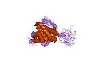
Bcl-2-associated X protein

Apoptosis regulator BAX also known as bcl-2-like protein 4 is a protein that in humans is encoded by the BAX gene.[1]
BAX is a member of the Bcl-2 gene family. Apoptosis regulator BAX promotes apoptosis by binding to and antagonizing the Bcl-2 protein.[1]
The BAX gene was the first identified pro-apoptotic member of the Bcl-2 protein family.[2] Bcl-2 family members share one or more of the four characteristic domains of homology entitled the Bcl-2 homology (BH) domains (named BH1, BH2, BH3 and BH4), and can form hetero- or homodimers.[2][3] Bcl-2 proteins act as anti- or pro-apoptotic regulators that are involved in a wide variety of cellular activities.
Orthologs of the BAX gene [4] have been identified in most mammals for which complete genome data are available. Certain members of the BCL-2 protein family, such as Bcl-2, Bcl-xl and Mcl-1 are anti-apoptotic, whilst others are pro-apoptotic. BAX is a pro-apoptotic Bcl-2 protein containing BH1, BH2 and BH3 domains.
Function
In healthy mammalian cells, the majority of BAX is found in the cytosol, but upon initiation of apoptotic signaling, Bax undergoes a conformational shift. Upon induction of apoptosis, BAX becomes organelle membrane-associated, and in particular, mitochondrial membrane associated.[5][6][7][8][9]
BAX is believed to interact with, and induce the opening of the mitochondrial voltage-dependent anion channel, VDAC.[10] Alternatively, growing evidence also suggests that activated BAX and/or Bak form an oligomeric pore, MAC in the outer membrane.[11] This results in the release of cytochrome c and other pro-apoptotic factors from the mitochondria, often referred to as mitochondrial outer membrane permeabilization, leading to activation of caspases.[12] This defines a direct role for BAX in mitochondrial outer membrane permeabilization, a role common to the Bcl-2 proteins containing the BH1, BH2 and BH3 domains.
Role in Cancer
The expression of BAX is upregulated by the tumor suppressor protein p53, and BAX has been shown to be involved in p53-mediated apoptosis.[13][14][15] The p53 protein is a transcription factor[13][14][15] that, when activated as part of the cell's response to stress, regulates many downstream target genes, including BAX. Wild-type p53 has been demonstrated to upregulate the transcription of a chimeric reporter plasmid utilizing the consensus promoter sequence of BAX approximately 50-fold over mutant p53.[13][14][15] Thus it is likely that p53 promotes BAX's apoptotic faculties in vivo as a primary transcription factor. However, p53 also has a transcription-independent role in apoptosis. In particular, p53 interacts with Bax, promoting its activation as well as its insertion into the mitochondrial membrane.[13][14][15]
Binding of HA-BAD to BCL-xL and concomitant disruption of BAX:BCL-xL interaction was found to partly reverse paclitaxel resistance in human ovarian cancer cells.[16]
Interactions
Bcl-2-associated X protein has been shown to interact with:
- Bcl-2,[2][3][17][18][19]
- BCL2L1,[3][16][20][21]
- BCL2A1[3][22]
- SH3GLB1,[8][23]
- SLC25A4,[24]
- VDAC1,[10][12] and
- YWHAQ.[25]
See also
- Apoptosis
- Apoptosome
- Bcl-2
- BH3 interacting domain death agonist (BID)
- Caspases
- Cytochrome c
- Noxa
- Mitochondrion
- p53 upregulated modulator of apoptosis (PUMA)
References
- ↑ 1.0 1.1 "Entrez Gene: BCL2-associated X protein".
- ↑ 2.0 2.1 2.2 Oltvai ZN, Milliman CL, Korsmeyer SJ (August 1993). "Bcl-2 heterodimerizes in vivo with a conserved homolog, Bax, that accelerates programmed cell death". Cell 74 (4): 609–19. doi:10.1016/0092-8674(93)90509-O. PMID 8358790.
- ↑ 3.0 3.1 3.2 3.3 Sedlak TW, Oltvai ZN, Yang E, Wang K, Boise LH, Thompson CB, Korsmeyer SJ (August 1995). "Multiple Bcl-2 family members demonstrate selective dimerizations with Bax". Proc. Natl. Acad. Sci. U.S.A. 92 (17): 7834–8. doi:10.1073/pnas.92.17.7834. PMC 41240. PMID 7644501.
- ↑ "OrthoMaM phylogenetic marker: BAX coding sequence".
- ↑ Gross A, Jockel J, Wei MC, Korsmeyer SJ (July 1998). "Enforced dimerization of BAX results in its translocation, mitochondrial dysfunction and apoptosis". EMBO J. 17 (14): 3878–85. doi:10.1093/emboj/17.14.3878. PMC 1170723. PMID 9670005.
- ↑ Hsu YT, Wolter KG, Youle RJ (April 1997). "Cytosol-to-membrane redistribution of Bax and Bcl-X(L) during apoptosis". Proc. Natl. Acad. Sci. U.S.A. 94 (8): 3668–72. doi:10.1073/pnas.94.8.3668. PMC 20498. PMID 9108035.
- ↑ Nechushtan A, Smith CL, Hsu YT, Youle RJ (May 1999). "Conformation of the Bax C-terminus regulates subcellular location and cell death". EMBO J. 18 (9): 2330–41. doi:10.1093/emboj/18.9.2330. PMC 1171316. PMID 10228148.
- ↑ 8.0 8.1 Pierrat B, Simonen M, Cueto M, Mestan J, Ferrigno P, Heim J (January 2001). "SH3GLB, a new endophilin-related protein family featuring an SH3 domain". Genomics 71 (2): 222–34. doi:10.1006/geno.2000.6378. PMID 11161816.
- ↑ Wolter KG, Hsu YT, Smith CL, Nechushtan A, Xi XG, Youle RJ (December 1997). "Movement of Bax from the cytosol to mitochondria during apoptosis". J. Cell Biol. 139 (5): 1281–92. doi:10.1083/jcb.139.5.1281. PMC 2140220. PMID 9382873.
- ↑ 10.0 10.1 Shi Y, Chen J, Weng C, Chen R, Zheng Y, Chen Q, Tang H (June 2003). "Identification of the protein-protein contact site and interaction mode of human VDAC1 with Bcl-2 family proteins". Biochem. Biophys. Res. Commun. 305 (4): 989–96. doi:10.1016/S0006-291X(03)00871-4. PMID 12767928.
- ↑ Buytaert E, Callewaert G, Vandenheede JR, Agostinis P (2006). "Deficiency in apoptotic effectors Bax and Bak reveals an autophagic cell death pathway initiated by photodamage to the endoplasmic reticulum". Autophagy 2 (3): 238–40. doi:10.4161/auto.2730. PMID 16874066.
- ↑ 12.0 12.1 Weng C, Li Y, Xu D, Shi Y, Tang H (March 2005). "Specific cleavage of Mcl-1 by caspase-3 in tumor necrosis factor-related apoptosis-inducing ligand (TRAIL)-induced apoptosis in Jurkat leukemia T cells". J. Biol. Chem. 280 (11): 10491–500. doi:10.1074/jbc.M412819200. PMID 15637055.
- ↑ 13.0 13.1 13.2 13.3 Miyashita T, Krajewski S, Krajewska M, Wang HG, Lin HK, Liebermann DA, Hoffman B, Reed JC (June 1994). "Tumor suppressor p53 is a regulator of bcl-2 and bax gene expression in vitro and in vivo". Oncogene 9 (6): 1799–805. PMID 8183579.
- ↑ 14.0 14.1 14.2 14.3 Selvakumaran M, Lin HK, Miyashita T, Wang HG, Krajewski S, Reed JC, Hoffman B, Liebermann D (June 1994). "Immediate early up-regulation of bax expression by p53 but not TGF beta 1: a paradigm for distinct apoptotic pathways". Oncogene 9 (6): 1791–8. PMID 8183578.
- ↑ 15.0 15.1 15.2 15.3 Miyashita T, Reed JC (January 1995). "Tumor suppressor p53 is a direct transcriptional activator of the human bax gene". Cell 80 (2): 293–9. doi:10.1016/0092-8674(95)90412-3. PMID 7834749.
- ↑ 16.0 16.1 Strobel T, Tai YT, Korsmeyer S, Cannistra SA (November 1998). "BAD partly reverses paclitaxel resistance in human ovarian cancer cells". Oncogene 17 (19): 2419–27. doi:10.1038/sj.onc.1202180. PMID 9824152.
- ↑ Hoetelmans RW (2004). "Nuclear partners of Bcl-2: Bax and PML". DNA Cell Biol. 23 (6): 351–4. doi:10.1089/104454904323145236. PMID 15231068.
- ↑ Lin B, Kolluri SK, Lin F, Liu W, Han YH, Cao X, Dawson MI, Reed JC, Zhang XK (2004). "Conversion of Bcl-2 from protector to killer by interaction with nuclear orphan receptor Nur77/TR3". Cell 116 (4): 527–40. doi:10.1016/S0092-8674(04)00162-X. PMID 14980220.
- ↑ Komatsu K, Miyashita T, Hang H, Hopkins KM, Zheng W, Cuddeback S, Yamada M, Lieberman HB, Wang HG (2000). "Human homologue of S. pombe Rad9 interacts with BCL-2/BCL-xL and promotes apoptosis". Nat. Cell Biol. 2 (1): 1–6. doi:10.1038/71316. PMID 10620799.
- ↑ Zhang H, Nimmer P, Rosenberg SH, Ng SC, Joseph M (2002). "Development of a high-throughput fluorescence polarization assay for Bcl-x(L)". Anal. Biochem. 307 (1): 70–5. doi:10.1016/S0003-2697(02)00028-3. PMID 12137781.
- ↑ Gillissen B, Essmann F, Graupner V, Stärck L, Radetzki S, Dörken B, Schulze-Osthoff K, Daniel PT (2003). "Induction of cell death by the BH3-only Bcl-2 homolog Nbk/Bik is mediated by an entirely Bax-dependent mitochondrial pathway". EMBO J. 22 (14): 3580–90. doi:10.1093/emboj/cdg343. PMC 165613. PMID 12853473.
- ↑ Zhang H, Cowan-Jacob SW, Simonen M, Greenhalf W, Heim J, Meyhack B (2000). "Structural basis of BFL-1 for its interaction with BAX and its anti-apoptotic action in mammalian and yeast cells". J. Biol. Chem. 275 (15): 11092–9. doi:10.1074/jbc.275.15.11092. PMID 10753914.
- ↑ Cuddeback SM, Yamaguchi H, Komatsu K, Miyashita T, Yamada M, Wu C, Singh S, Wang HG (2001). "Molecular cloning and characterization of Bif-1. A novel Src homology 3 domain-containing protein that associates with Bax". J. Biol. Chem. 276 (23): 20559–65. doi:10.1074/jbc.M101527200. PMID 11259440.
- ↑ Marzo I, Brenner C, Zamzami N, Jürgensmeier JM, Susin SA, Vieira HL, Prévost MC, Xie Z, Matsuyama S, Reed JC, Kroemer G (1998). "Bax and adenine nucleotide translocator cooperate in the mitochondrial control of apoptosis". Science 281 (5385): 2027–31. Bibcode:1998Sci...281.2027M. doi:10.1126/science.281.5385.2027. PMID 9748162.
- ↑ Nomura M, Shimizu S, Sugiyama T, Narita M, Ito T, Matsuda H, Tsujimoto Y (2003). "14-3-3 Interacts directly with and negatively regulates pro-apoptotic Bax". J. Biol. Chem. 278 (3): 2058–65. doi:10.1074/jbc.M207880200. PMID 12426317.
| |||||||||
| ||||||||||||||||||||||||||||||||||||||||||||||