Arthrogryposis
| Arthrogryposis | |
|---|---|
.png) Drawing of an infant with arthrogryposis | |
| Classification and external resources | |
| ICD-10 | Q74.3 |
| ICD-9 | 728.3, 754.89 |
| OMIM | 108110 108120 208100 301830 601701 208200 108200 301830 208155 601680 108145 208085 |
| DiseasesDB | 31688 31816 |
| eMedicine | ped/142 |
| MeSH | D001176 |
Arthrogryposis multiplex congenita (AMC), or simply arthrogryposis, describes congenital joint contractures in two or more areas of the body. It derives its name from Greek, literally meaning "curving of joints" (arthron, "joint"; grȳpōsis, late Latin form of late Greek grūpōsis, "hooking").[1] Children born with one or more joint contractures have abnormal fibrosis of the muscle tissue causing muscle shortening, and therefore are unable to do passive extension and flexion in the affected joint or joints.[2] AMC has been divided into three groups: amyoplasia, distal arthrogryposis, and syndromic. Amyoplasia is characterized by severe joint contractures and muscle weakness.[3] Distal arthrogryposis mainly involves the hands and feet. Types of arthrogryposis with a primary neurological or muscle disease belong to the syndromic group.[3]
Epidemiology
Arthrogryposis is a rare condition. Some authors say the overall prevalence is one in 3000[3] and others say it is one in 11000-12000 among European live births.[4] Congenital clubfoot is the most common single contracture and its prevalence is one in 500 live births.[3]
Classification
Some of the different types of AMC include:
- Arthrogryposis multiplex due to muscular dystrophy.[5][6]
- Arthrogryposis ectodermal dysplasia other anomalies, also known as Cote Adamopoulos Pantelakis syndrome, Trichooculodermovertebral syndrome, TODV syndrome and Alves syndrome.[7][8]
- Arthrogryposis epileptic seizures migrational brain disorder.[9]
- Arthrogryposis IUGR thoracic dystrophy,also known as Van Bervliet syndrome.[10][11]
- Arthrogryposis like disorder, also known as Kuskokwim disease.[12]
- Arthrogryposis-like hand anomaly and sensorineural deafness.[13][14]
- Arthrogryposis multiplex congenita CNS calcification.[15]
- Arthrogryposis multiplex congenita distal (AMCD), also known as X-linked spinal muscular atrophy type 2[16][17][18]
- Gordon Syndrome, also known as Distal Arthrogryposis, Type 2A.[19]
- Arthrogryposis multiplex congenita, distal type 2B, also known as Freeman-Sheldon syndrome variant.[20]
- Arthrogryposis multiplex congenita neurogenic type (AMCN).[21] This particular type of AMC has been linked to the AMCN gene on locus 5q35.[22][23] Arthrogryposis multiplex congenita pulmonary hypoplasia, also with a large number of synonyms.[24][25]
- Arthrogryposis multiplex congenita whistling face, also known as Illum syndrome.[26][27][28][29]
- Arthrogryposis multiplex congenita, distal type 1 (AMCD1).[30]
- Arthrogryposis ophthalmoplegia retinopathy, also known as Oculomelic amyoplasia.[31][32][33]
- Arthrogryposis renal dysfunction cholestasis syndrome, also known as ARC Syndrome.[34][35]
Signs and Symptoms
Almost every joint in a patient with arthrogryposis is affected, because in 84% all limbs are involved, in 11% only the legs and in 4% only the arms are involved.[36] Every joint in the body has typical signs and symptoms like the shoulder (internal rotation), wrist (volar and ulnar), hand (fingers in fixed flexion and thumb in palm), hip (flexed, abducted and externally rotated, frequently dislocated), elbow (extension and pronation) and foot (clubfoot).[2] The range of motion capability can be different between joints because of the different deviations.[37] Some types of arthrogryposis like amyoplasia have a symmetrical joint/limb involvement, with normal sensations.[36][37] The contractures in the joints are sometimes resulting in a reduced walking development in the first 5 years.[37] The intelligence is normal to above normal in children with amyoplasia.[36] But it is unknown how many of these children have an above normal intelligent and there is no literature available about the cause of this symptom. There are a few syndromes like the Freeman-Sheldon and Gordon syndrome, which have craniofacial involvement.[36] The amyoplasia form of arthrogryposis is sometimes accompanied with a midline facial hemangioma.[36] Arthrogryposis is a not a diagnosis but a clinical finding. So this disease is often accompanied with other syndromes or diseases. These other diagnosis can be found in every single organ in a patient. There are a few slightly more common diagnoses such as pulmonary hypoplasia, cryptorchidism, congenital heart defects, tracheoesophageal fistulas, inguinal hernias, cleft palate, and eye abnormalities.[38]
Causes
Research of arthrogryposis has shown that anything that inhibits normal joint movement before birth can result in joint contractures.[3] Arthrogryposis could be caused by genetic and environmental factors. In principle: any factor that entails fetal movement of the fetus can result to congenital contractures.[36] The exact causes of arthrogryposis are unknown yet.
Extrinsic factors
The malformations of arthrogryposis can be secondary to environmental factors such as: decreased intrauterine movement, oligohydramnios (low volume or abnormal distribution of intrauterine fluid), and defects in the fetal blood supply. Other causes could be: hyperthermia, limb immobilization and viral infections. Myasthenia gravis of the mother leads also in rare cases to arthrogryposis. The major cause in humans is fetal akinesia.[2] However, this is disputed lately.[2]
Intrinsic Factors
Arthrogryposis could also be caused by intrinsic factors. Think of molecular, muscle- and connective tissues development disorders or neurological abnormalities.
Molecular basis
Research has shown that there are more than 35 specific genetic disorders associated with arthrogryposis. Most of those mutations are missense, which means the mutation results in a different amino acid. Other mutations that could cause arthrogryposis are: single gene defects (X-linked recessive, autosomal recessive and autosomal dominant), mitochondrial defects and chromosomal disorders (for example: trisomy 18).[36] This is mostly seen in distal arthrogryposis. Mutations in at least five genes (TNN12, TNNT3, TPM2, MYH3 and MYH8) could cause distal arthrogryposis.[3] There could be also connective tissue-, neurological of muscle development disorders.[3]
Muscle- and connective tissue development disorders
Loss of muscle mass with an imbalance of muscle power at the joint can lead to connective tissue abnormality.[2] This leads to joint fixation and reduced fetal movement.[2] Also muscle abnormalities could lead to reduce of fetal movement. Those could be: dystrophy, myopathy and mitochondrial disorders. This is mostly the result of abnormal function of the dystrophin-glycoprotein-associated complex in the sarcolemma of skeletal muscles.[2]
Neurological abnormalities.
70-80% of the cases of arthrogryposis are caused by neurological abnormalities. Most of these are underlying of a genetic syndrome or the result of environmental factors.[3]
The underlying aetiology and pathogenesis of congenital contractures, particularly arthrogryposis and the mechanism of the mutations remains an active area of investigation. Because identifying these factors could help to develop treatment and congenital finding of arthrogryposis.[3][39]
Diagnosis
There is currently a lot of research on prenatal diagnosis. The diagnosis can be made prenatally in approximately 50% of fetuses presenting arthrogryposis.[40] It could be found during routine ultrasound scanning showing a lack of mobility and abnormal position of the foetus.[2] Nowadays there are more options for visualization of details and structures can be seen well, like the use of 4D ultrasound.[2] In clinic a child can be diagnosed with arthrogryposis with physical examination, confirmed by ultrasound, MRI,[3][40] or muscle biopsy.[40]
Treatment
The treatment of arthrogryposis includes physical therapy, splinting and surgery.[3] The primary long-term goals of these treatments are increasing joint mobility, muscle strength and the development of adaptive use patterns that allow for walking and independence with activities of daily living. Since arthrogryposis includes many different types, the treatment varies between patients depending on the symptoms.[3] Only a few good articles exist in which a surgical technique that is used to treat arthrogryposis is described. These surgeries are explained below.
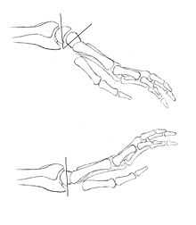
Wrist surgery in Arthrogryposis
Children with the amyoplasia type of arthrogryposis usually have flexed and ulnarly deviated wrists.[2] Dorsal carpal wedge osteotomy is indicated for wrists with excessive flexion contracture deformity when non-surgical interventions such as occupational therapy and splinting have failed to improve function. On the dorsal side, at the level of the midcarpus, a wedge osteotomy is made. Sufficient bone is resected to at least be able to put the wrist in a neutral position. If the wrist is also ulnarly deviated, more bone can be taken from the radial side to correct this abnormality. This position is held into place with two cross K-wires. In addition, a tendon transfer of the extensor carpi ulnaris to the extensor carpi radialis brevis may be performed to correct ulnar deviation or wrist extension weakness, or both. This tendon transfer is only used if the extensor carpi ulnaris appears to be functional enough.[41]
Thumb surgery in Arthrogryposis
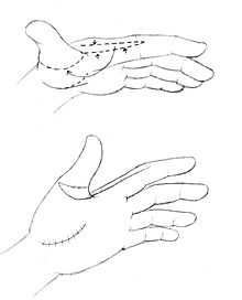
The soft tissue envelope in congenital contractual conditions such as clasped or arthrogrypotic thumbs is often deficient in two planes, the thumb-index web and the flexor aspect of the thumb. There is often an appearance of increased skin at the base of the index finger that is part of the deformity. This tissue can be used to resurface the thumb-index web after a comprehensive release of all the tight structures to allow for a larger range of motion of the thumb. This technique is called the index rotation flap. The flap is taken from the radial side of the index finger. It is proximally based at the distal edge of the thumb-index web. The flap is made as wide as possible, but still small enough to close with the excessive skin on the palmar side of the index finger. The flap is rotated around the tightest part of the thumb to the metacarpophalangeal joint of the thumb, allowing for a larger range of motion.[42]
Other surgeries
Many other surgeries are also able to improve function in joints of arthrogryposis patients. These surgeries usually exist out of tendon transfers and skin flap movements, adjusted to the individual.
Prognosis
There is still little literature available about the prognosis of patient with arthrogryposis. The search for a prognostic factor is difficult, because of the small number of patients. But there are a few factors that seem to have an influence on the prognosis like the progressive muscle activity improvement. Results of a study showed an increase in strength during growth after the recovery of a passive range of motion in a useful arc.[37] Other positive prognostic factors for independent walking were active hips and knees, hip flexion contractures of less than 20 degrees and knee flexion contractures less than 15 degrees without severe scoliosis.[37]
See also
References
- ↑ "The Free Dictionary: Arthrogryposis". Retrieved 11 July 2013.
- ↑ 2.0 2.1 2.2 2.3 2.4 2.5 2.6 2.7 2.8 2.9 Kalampokas, Emmanouil; Kalampokas, Theodoros; Sofoudis, Chrisostomos; Deligeoroglou, Efthymios; Botsis, Dimitrios (2012). "Diagnosing Arthrogryposis Multiplex Congenita: A Review". ISRN Obstetrics and Gynecology 2012: 1. doi:10.5402/2012/264918. PMC 3461621. PMID 23050160.
- ↑ 3.0 3.1 3.2 3.3 3.4 3.5 3.6 3.7 3.8 3.9 3.10 3.11 Bamshad, Michael; Van Heest, AE; Pleasure, D (2009). "Arthrogryposis: A Review and Update". The Journal of Bone and Joint Surgery (American) 91 (Suppl 4): 40–6. doi:10.2106/JBJS.I.00281. PMC 2698792. PMID 19571066.
- ↑ Hoff, Jana Midelfart; Loane, Maria; Gilhus, Nils Erik; Rasmussen, Svein; Daltveit, Anne Kjersti (2011). "Arthrogryposis multiplexa congenita: An epidemiologic study of nearly 9 million births in 24 EUROCAT registers". European Journal of Obstetrics & Gynecology and Reproductive Biology 159 (2): 347–50. doi:10.1016/j.ejogrb.2011.09.027. PMID 22005589.
- ↑ Smit, L. M. E.; Earth, P. G. (2008). "Arthrogryposis Multiplex Congenita due to Congenital Myasthenia". Developmental Medicine & Child Neurology 22 (3): 371–4. doi:10.1111/j.1469-8749.1980.tb03718.x. PMID 6446471.
- ↑ Banker, Betty Q.; Victor, Maurice; Adams, Raymond D. (1957). "Arthrogryposis Multiplex Due to Congenital Muscular Dystrophy". Brain 80 (3): 319–34. doi:10.1093/brain/80.3.319. PMID 13471804.
- ↑ Arthrogryposis and ectodermal dysplasia at NIH's Office of Rare Diseases
- ↑ Stoll, C; Alembik, Y; Finck, S; Janser, B (1992). "Arthrogryposis, ectodermal dysplasia and other anomalies in two sisters". Genetic counseling 3 (1): 35–9. PMID 1590979.
- ↑ "ORPHA1139 Arthrogryposis - epileptic seizures - migrational brain disorder". Orphanet.ORPHANET - About rare diseases - About orphan drugs
- ↑ ORPHANET - About rare diseases - About orphan drugs
- ↑ Arthrogryposis IUGR thoracic dystrophy at NIH's Office of Rare Diseases
- ↑ CTD: Disease Not Found
- ↑ CTD: Disease Not Found
- ↑ Arthrogryposis-like hand anomaly and sensorineural deafness at NIH's Office of Rare Diseases
- ↑ Arthrogryposis multiplex congenita CNS calcification at NIH's Office of Rare Diseases
- ↑ Online 'Mendelian Inheritance in Man' (OMIM) 301830
- ↑ ORPHANET - About rare diseases - About orphan drugs
- ↑ Kizilates, Sevim Ünal; Talim, Beril; Sel, Kutay; Köse, Gulsen; Caglar, Melda (2005). "Severe lethal spinal muscular atrophy variant with arthrogryposis". Pediatric Neurology 32 (3): 201–4. doi:10.1016/j.pediatrneurol.2004.10.003. PMID 15730903. INIST:16634238.
- ↑ Gordon Syndrome
- ↑ http://www.medinet.lk/journals/CMJ/2001/december/arthrogryposis.htm
- ↑ Arthrogryposis Multiplex Congenita, Neurogenic Type - What does AMCN stand for? Acronyms and abbreviations by the Free Online Dictionary
- ↑ CTD: Disease Not Found
- ↑ CTD: Disease Not Found
- ↑ ORPHANET - About rare diseases - About orphan drugs
- ↑ Leichtman, Lawrence G.; Say, Burhan; Barber, Nancy (1980). "Primary pulmonary hypoplasia and arthrogryposis multiplex congenita". The Journal of Pediatrics 96 (5): 950–1. doi:10.1016/S0022-3476(80)80591-9. PMID 7365612.
- ↑ Illum, N.; Reske-Nielsen, Edith; Skovby, F.; Askjaer, S.; Bernsen, Alice (2008). "Lethal Autosomal Recessive Arthrogryposis Multiplex Congenita with Whistling Face and Calcifications of the Nervous System". Neuropediatrics 19 (4): 186–92. doi:10.1055/s-2008-1052443. PMID 3205375.
- ↑ CTD: Disease Not Found
- ↑ ORPHANET - About rare diseases - About orphan drugs
- ↑ Arthrogryposis multiplex congenita whistling face at NIH's Office of Rare Diseases
- ↑ Arthrogryposis multiplex congenita at NIH's Office of Rare Diseases
- ↑ ORPHANET - About rare diseases - About orphan drugs
- ↑ Arthrogryposis ophthalmoplegia retinopathy at NIH's Office of Rare Diseases
- ↑ Schrander-Stumpel, C T; Höweler, C J; Reekers, A D; De Smet, N M; Hall, J G; Fryns, J P (1993). "Arthrogryposis, ophthalmoplegia, and retinopathy: Confirmation of a new type of arthrogryposis". Journal of Medical Genetics 30 (1): 78–80. doi:10.1136/jmg.30.1.78. PMC 1016242. PMID 8423615.
- ↑ Rocco, M.; Callea, F.; Pollice, B.; Faraci, M.; Campiani, F.; Borrone, C. (1995). "Arthrogryposis, renal dysfunction and cholestasis syndrome: Report of five patients from three Italian families". European Journal of Pediatrics 154 (10): 835–9. doi:10.1007/BF01959793. PMID 8529684.
- ↑ Arthrogryposis renal dysfunction cholestasis syndrome at NIH's Office of Rare Diseases
- ↑ 36.0 36.1 36.2 36.3 36.4 36.5 36.6 Bevan, Wesley P.; Hall, Judith G.; Bamshad, Micheal; Staheli, Lynn T.; Jaffe, Kenneth M.; Song, Kit (2007). "Arthrogryposis Multiplex Congenita (Amyoplasia)". Journal of Pediatric Orthopaedics 27 (5): 594–600. doi:10.1097/BPO.0b013e318070cc76. PMID 17585274.
- ↑ 37.0 37.1 37.2 37.3 37.4 Fassier, Alice; Wicart, Philippe; Dubousset, Jean; Seringe, Raphaël (2009). "Arthrogryposis multiplex congenita. Long-term follow-up from birth until skeletal maturity". Journal of Children's Orthopaedics 3 (5): 383–90. doi:10.1007/s11832-009-0187-4. PMC 2758174. PMID 19669823.
- ↑ Brooks, James G; Coster, Douglas J (1994). "Arthrogryposis multiplex congenita: A report of two cases". Australian and New Zealand Journal of Ophthalmology 22 (2): 127–32. doi:10.1111/j.1442-9071.1994.tb00780.x. PMID 7917267.
- ↑ Wynne-Davies, R; Williams, PF; O'Connor, JC (1981). "The 1960s epidemic of arthrogryposis multiplex congenita: A survey from the United Kingdom, Australia and the United States of America". The Journal of bone and joint surgery. British volume 63–B (1): 76–82. PMID 7204479.
- ↑ 40.0 40.1 40.2 Rink, Britton D. (2011). "Arthrogryposis: A Review and Approach to Prenatal Diagnosis". Obstetrical & Gynecological Survey 66 (6): 369–77. doi:10.1097/OGX.0b013e31822bf5bb. PMID 21851751.
- ↑ Van Heest, Ann E.; Rodriguez, Rudy (2013). "Dorsal Carpal Wedge Osteotomy in the Arthrogrypotic Wrist". The Journal of Hand Surgery 38 (2): 265–70. doi:10.1016/j.jhsa.2012.10.034. PMID 23267756.
- ↑ Ezaki, Marybeth; Oishi, Scott N. (2010). "Index Rotation Flap for Palmar Thumb Release in Arthrogryposis". Techniques in Hand & Upper Extremity Surgery 14 (1): 38–40. doi:10.1097/BTH.0b013e3181d44583. PMID 20216051.
External links
- AMC Support (United States)
- Avenues (United States)
- Project Scissor Gait Foundation
- The Michael Fund (United States)
- Women for AMC Awareness
- The Arthrogryposis Group (United Kingdom)
- Enabled - A True Story About A Lady with Arthrogryposis (United Kingdom)
| ||||||||||||||||||||||||||||||||||||||||
| ||||||||||||||||||||||||||||||||||||||||||||||||||||||||||||||||||||||||||||||||||||||||||