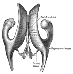
Anterior horn of lateral ventricle
| Anterior horn of lateral ventricle | |
|---|---|
 Drawing of a cast of the ventricular cavities, viewed from above. | |
 Drawing of a cast of the ventricular cavities, viewed from the side. | |
| Details | |
| Latin | Cornu anterius |
| Identifiers | |
| Gray's | p.829 |
| NeuroNames | hier-202 |
| NeuroLex ID | Anterior horn of lateral ventricle |
| TA | A14.1.09.273 |
| FMA | 74520 |
| Anatomical terms of neuroanatomy | |
The anterior horn of the lateral ventricle (also anterior cornu of the lateral ventricle, frontal horn of the lateral ventricle or precornu) is a portion of the lateral ventricle that passes forward and laterally, with a slight inclination downward, from the Interventricular foramen into the frontal lobe, curving around the anterior end of the caudate nucleus. Its floor is formed by the upper surface of the reflected portion of the corpus callosum, the rostrum. It is bounded medially by the anterior portion of the septum pellucidum, and laterally by the head of the caudate nucleus. Its apex reaches the posterior surface of the genu of the corpus callosum.
Additional images
-
Human brain right dissected lateral view
-
Ventricles of braian and basal ganglia.Superior view. Horizontal section.Deep dissection
-
Ventricles of brain and basal ganglia.Superior view. Horizontal section.Deep dissection
References
This article incorporates text in the public domain from the 20th edition of Gray's Anatomy (1918)
External links
- Atlas image: n1a4p2 at the University of Michigan Health System
- Atlas image: n1a4p5 at the University of Michigan Health System
- http://www2.umdnj.edu/~neuro/studyaid/Practical2000/Q42.htm
| ||||||||||||||||||||||||||||


