Anterior cruciate ligament reconstruction
| Anterior cruciate ligament reconstruction | |
|---|---|
| Intervention | |
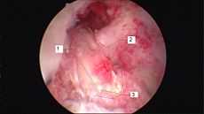 Arthroscopic anterior cruciate ligament (ACL) reconstruction (right knee). The tendon of the semitendinosus muscle was prelevated, folded and used as an autograft (1). It appears through the remnant of the injured original ACL (3). The autograft then courses upwardly and backwardly in front of the posterior cruciate ligament (2). | |
| ICD-9-CM | 81.45 |
| MedlinePlus | 007208 |
Anterior cruciate ligament reconstruction (ACL reconstruction) is a surgical tissue graft replacement of the anterior cruciate ligament, located in the knee, to restore its function after anterior cruciate ligament injury. The torn ligament is removed from the knee before the graft is inserted. The surgery is performed arthroscopically.
Types of grafts

Two alternative sources of replacement material for ACL reconstruction are commonly utilized:
- Autografts (employing bone or tissue harvested from the patient's body), and
- Allografts (using bone or tissue from a donor's body, typically a cadaver's or a live donor).
Since the tissue is one's own in an autograft the probability of rejection (sans infection) is minimal.
Sterilization and redundant donor screening process make allografts a generally safe choice for patients; however, risks remain. Irradiation of donor content to remove infectious agents potentially weakens the selected tendon, although for ACL surgery the weakened tendon is generally as strong as the replaced ligament.[1] Infection may also require removal of the graft.
- Synthetic tissue suitable to ACL reconstruction has also been developed. Few data exist on its strength or reliability.
Allograft
The patellar tendon, anterior tibialis tendon, or Achilles tendon may be recovered from a cadaver and used as an allograft in reconstruction. The Achilles tendon, due to its large size, must be shaved to fit within the joint cavity. There is a slight chance of rejection, which would lead to more surgery to remove the graft and replace it.
Autograft
An accessory hamstring or part of the patellar tendon are the most common donor tissues used in autografts. Quadriceps tendon can also be used if the patellar tendon has been harvested in a previous surgery.
Hamstring tendon
Hamstring autografts are made with the semitendinosus tendon either alone, or accompanied by the gracilis tendon for a stronger graft. The semitendinosus is an accessory hamstring (the primary hamstrings are left intact), and the gracilis is actually not a hamstring, but an accessory adductor (the primary adductors are left intact as well). The two tendons are commonly combined and referred to as a four strand hamstring graft, made by a long piece (about 25 cm) which is removed from each tendon. The tendon segments are folded and braided together to form a quadruple thickness strand for the replacement graft. The braided segment is threaded through the heads of tibia and femur and its ends fixated with screws on the opposite sides of the two bones.
Unlike the patellar tendon, the hamstring tendon's fixation to the bone can be affected by motion in the post-operative phase. Therefore, following surgery, a brace is often used to immobilize the knee for one to two weeks while the most critical healing takes place. Evidence suggests that the hamstring tendon graft does just as well, or nearly as well, as the patellar tendon graft in the long-term[2].
Many patients struggle with recovery after a hamstring graft procedure. Problems include strengthening of the quadriceps, T-band and calf. Proper healing procedures and medical care (physical therapy) are essential to regain strength.
The main surgical wound is over the upper proximal tibia, avoiding the typical pain sensation when one kneels down. The wound is typically smaller than the patellar tendon graft and hence less pain after the operation. A new technique for minimal-invasive harvesting from the back of the knee has been developed in the last years. This technique is faster, easier and produces a significantly smaller wound.[3] Unfortunately, this less invasive procedure can still cause nerve damage around the scar and throughout the distal portion of the leg. In an effort to fix this issue, surgeons have tested different incision directions in an effort to decrease nerve damage and sensory loss near the incision site. A study published in Knee Surgery Sports Traumatology Arthroscopy (Sabat and Kumar, 2012, p. 2093) about whether vertical, transverse or oblique cuts result in the least amount of nerve and sensory damage came to the conclusion that, “With the oblique incision, the direction of incision is almost parallel to the infrapatella branch of the saphenous nerve, thus the risk of injury to that nerve is the lowest.”
There seems to be some controversy as to how well a hamstring tendon regenerates after the harvesting. Most studies suggest that the tendon can be regenerated at least partially, while still being inferior in strength to the original tendon.[4][5]
Advantages of using a hamstring graft includes its high “load to failure” strength, the stiffness of the graft, and the low postoperative morbidity. It has shown that the natural ACL can withstand a load of up to 2160 Newtons.By using a hamstring graft, this number doubles. The "load to failure" is twice as high as the natural ACL, helping to decrease risk of becoming injured again. The stiffness of a hamstring graft also contributes to this risk factor. If the graft is stretching out instead of staying stiff, this increases the shift between the tibia and the fibula. In comparison to the stiffness of the natural ACL, the hamstring graft quadruples (Bartlett, Clatworthy and Ngugen, 2001). Therefore, using a hamstring graft for a torn ACL has a variety of benefits including stability and recovery purposes, decreasing the chance of re-injury.
Patellar tendon
The patellar tendon connects the patella (kneecap) to the tibia (shin). The graft is taken from the injured knee, but in some circumstances, such as a second operation, the other knee may be used. The middle third of the tendon is used, with bone fragments removed on each end. The graft is then threaded through holes drilled in the tibia and femur, and finally screwed into place.
The graft is slightly larger than a hamstring graft, however graft size is not a determinant of outcome. The most important factor in determining the outcome is correct graft placement.
The disadvantages include: 1. Increased wound pain. 2. Increased scar formation as compared to a hamstring tendon operation. 3. Risk of fracturing the patella during harvesting of the graft. 4. Increased risk of tendinitis. 5. Increased levels of pain with activities that require kneeling years after post op.[6] In general, some or all of these disadvantages could be attributable to the phenomenon of post-operative patellar tendon shortening.[7]
Choice of graft
Type
Typically, age and lifestyle choices help decide the type of graft to be used for ACL reconstruction.[6] The overall factors in knee stability are correct graft placement by the surgeon and treatment of other menisco-ligament injuries in the knee, rather than type of graft.
The advantages of an allograft are that the patient does not sustain additional injury through removal of a tendon, thus improving recovery time, and that total procedure time is slightly reduced. Disadvantages are the risk of infection from foreign bodily materials and perhaps a slightly weaker graft.[8]
Site
No ideal graft site for ACL reconstruction exists. Surgeons have historically regarded patellar tendon grafts as the "gold standard" for knee stability,[6] however the procedure suffers a slightly higher complication rate, including knee pain when lunging.[9]
Hamstring grafts historically had problems with fixation slippage and stretching out over time. Modern fixation methods avoid graft slippage produce similarly stable outcomes with easier rehabilitation, less anterior knee pain and less joint stiffness.
Although there is generally less experience with the use of tibialis anterior grafts, preliminary data has shown no difference in short-term subjective outcomes between tibialis anterior allografts and patellar tendon allografts.[10]
Stem Cell Treatment during ACL Surgery
Recently, autologous stem cell transplantation using mesenchymal stem cells (MSCs) has been used to improve recovery time from ACL surgery, especially for athletes.[11] MSCs are multipotent stem cells, meaning they can differentiate into multiple cell types. In the case of mesenchymal stem cells, these cell types include osteoblasts (bone cells), adipocytes (fat cells), or chondrocytes (cartilage cells). Ligament tissue mainly consists of fibroblasts and extracellular matrix. The fibroblast is the main cell type but the extracellular matrix is made of collagen with proteoglycans. These fibroblasts are in charge of maintaining and regenerating new tissue. When introducing MSCs to induce new ligament growth, there are several factors that a tissue engineer must consider.[11] Ligament cells differ in size, respond to different cues in the cell environment, and express different cell surface markers, limiting the number of clinical treatments for accelerated repair of ACL tissue to MSCs and primary fibroblasts obtained from other ACL tissue. According to a study performed by Ge et al., the proliferation rate and collagen production were greater with MSCs than fibroblasts.[12] Therefore, most modern stem cell injections use MSCs to promote faster repair of the ACL and allow people such as athletes to return to their previous form faster than ever before.
In order for MSCs to differentiate into an ACL, they must be placed in a proper scaffold on which to grow and also must be in a bioreactor which will maintain a normal physiological environment for the cells to reproduce and proliferate effectively.[13] The scaffold must have the mechanical properties of a healthy ACL to sustain the ligament while it is in its primary form and maintain normal knee movement. It must also be biodegradable so that as the ligament grows, the scaffold degrades proportionally and eventually disappears altogether when the ligament has physiological mechanical properties.[14] Scaffolds that are used for ACL growth include collagen, silk, gelatin, poly-lactic acid, and glycosaminoglycins (GAGs).[15] Mechanical properties of the scaffolds are further enhanced through braiding and twisting of the scaffold materials.
The bioreactor is the environment in which the ACL will grow. It has to have similar properties to a knee joint so that when it is inserted into the body, it is not reject as foreign which could cause infection. Therefore, it has to have compatible pH levels, oxygen concentration levels, metabolite levels, and temperature as well as be sterile.[16]
Recovery
Initial physical therapy consists of range of motion (ROM) exercises, often with the guidance of a physical therapist. Range of motion exercises are used to regain the flexibility of the ligament, prevent or break down scar tissue from forming and reduce loss of muscle tone. Range of motion exercise examples include: quadriceps contractions and straight leg raises. In some cases, a continuous passive motion (CPM) device is used immediately after surgery to help with flexibility. The preferred method of preventing muscle loss is isometric exercises that put zero strain on the knee. Knee extension within two weeks is important with many rehab guidelines.
Approximately six weeks is required for the bone to attach to the graft. However, the patient can typically walk on their own and perform simple physical tasks prior to this with caution, relying on the surgical fixation of the graft until true healing (graft attachment to bone) has taken place. At this stage the first round of physical therapy can begin. This usually consists of careful exercises to regain flexibility and small amounts of strength. One of the more important benchmarks in recovery is the twelve weeks post-surgery period. After this, the patient can typically begin a more aggressive regimen of exercises involving stress on the knee, and increasing resistance. Jogging may be incorporated as well.
After four months, more intense activities such as running are possible without risk. After five months, light ball work may commence as the ligament is nearly regenerated. After six months, the reconstructed ACL is generally at full strength (ligament tissue has fully regrown), and the patient may return to activities involving cutting and twisting if a brace is worn. Recovery varies highly from case to case, and sometimes resumption of stressful activities may take a year or longer.
The reconstructed ACL has a high success rate. Studies show that cases in which the ACL retears are generally caused by a traumatic impact. Some studies indicate that wearing a brace during athletic activity does not reduce probability of re-injury to the ACL, but a study of active post-ACL replacement skiers shows a 64% reduction in re-injury likelihood by using a knee brace after recovery.[17] A sufficiently traumatic impact to retear the ACL is unlikely to be mitigated by the use of a brace.
Recovery Progression
Recovery is a four phase progression.
Phase 1 (0-2 weeks)
The goals of this phase are to:
- Eliminate swelling due to inactivity
- Progress from partial weight bearing to full weight bearing exercises
- Regain normal range of motion
- Increase quadriceps strength
- Increase hamstring strength
Some equipment that can be used and exercises that can be performed are:
- Use of Cryo-cuff
- - provides cold compression
- Isometric Contraction of Quads
- Quad Sets
- - stand against wall, push extended knee against rolled towel
- - progress to straight leg raised to 30deg.
- Wall Slides
- - To increase knee flexion
- Assisted Knee Flexion
- Towel Squeeze
- - Sit in chair, squeeze rolled towel between knees for 5 seconds. Relax & repeat.
- VMO Strengthening Exercise
- Supported Bilateral Calf-Raises
- walk without crutches
Phase 2 (2-12 weeks)
The goals of this phase are to:
- Regain full knee extension
- Restore knee flexion to +130°
- Perform a full squat properly
- Regain good balance and control
- Reestablish proper gait
Some exercises that can be performed are:
- Mini squats
- - Progress to full squats → single-leg half squat
- Mini Lunges
- - Progress to full lunges
- Leg Press
- - Double-leg → single
- Step-ups
- - lateral & forward
- Bridges
- - Double-leg → single
- - Floor → Swiss ball
- Hip Abduction w/ Theraband
- Hip Extension w/ Theraband
- Wobble board
- - Assisted → un-assisted → eyes closed (assisted → unassisted)
- Stork Stand
- - Assisted → un-assisted → eyes closed (assisted → unassisted) → unstable surface
- Static Proprioceptive hold/ball throwing
- Functional Exercises that can be performed at this time include:
- - Walking
- - Bike
- - Roman Chair
Phase 3 (3-6 months)
The goals of this phase are to:
- Regain full range of motion
- Regain full strength and power
- Increase agility
- allows for adaption to direction change, acceleration and deceleration
- Be able to perform restricted sports-specific drills
- Begin plyometric drills
Some exercises that can be performed are:
- Continue exercises from Phase 2, progress as necessary
- Jump & Land drills
- - Jump from block & stick landing
- - Double-leg landing → single-leg
- Plyometric Drills
- - Jumping over blocks, sideways & forward
- - Hopping up & down steps/stairs
Phase 4 (6-15 months)
The goal of this phase is a return to activity, however it requires an ability to perform some functional performance tests such as:
- Agility Tests
- Illinois Agility Test
- Zig Zag Agility Test
- These tests are used to test the ability of the knee to withstand cutting and planting maneuvers
- Standing Vertical Jump
- Here you jump straight in the air from a standing start and land on two feet as stable as possible.
- Heiden Hop Test
- Here you essentially jump as far as possible with the uninjured leg and land on the injured leg. Your ability to stick the landing is indicative of good knee function.
- Isokinetic Testing
- This is used to evaluate muscle strength.
- The individual should have at least 90% quadricep strength of the uninjured leg
- They should also have equal hamstring strength to their uninjured leg as well
Cost and logistics of the procedure
The cost of ACL surgery in the US is an unfortunate reality which affects whether or not a patient proceeds with the operation. The average out-of-pocket cost of ACL reconstruction is $2,339.43, according to a 2010 survey of ACL surgery patients.[18] Insurance companies may or may not cover the various billable components of ACL reconstruction, which may include: pre-op appointments, pre-op physical therapy, ACL reconstruction by the surgeon, an assistant's charge, anesthesia, the hospital or facility fee, rental fees for cryotherapy, prescriptions, transportation, crutches or wheel chair fees, leg brace fees, post op visits (such as to remove drain plugs and monitor swelling), and a physical therapy rehab program.
Despite the complexity of the procedure and numerous doctor visits involved, 80% - 90% of patients who have had the surgery said they had favorable results.[19]
References
- ↑ ACL (Anterior Cruciate Ligament) Graft Choices
- ↑ Pinczewski, Lyman, Salmon, Russell, Roe, Linklater, Leo, Jeffrey, Lucy, Vivianne, Justin, James (2007). "A 10-Year Comparison of Anterior Cruciate Ligament Reconstructions With Hamstring Tendon and Patellar Tendon Autograft". The American Journal of Sports Medicine. doi:10.1177/0363546506296042. Retrieved 2015-04-13.
- ↑ Franz W, Ulbrich J (2004). "A new technique for harvesting the semitendinosus tendon for cruciate ligament reconstruction". Arthroskopie 17: 104–7. doi:10.1007/s00142-004-0255-1.
- ↑ Okahashi K, Sugimoto K, Iwai M et al. (June 2006). "Regeneration of the hamstring tendons after harvesting for arthroscopic anterior cruciate ligament reconstruction: a histological study in 11 patients". Knee Surg Sports Traumatol Arthrosc 14 (6): 542–5. doi:10.1007/s00167-006-0068-z. PMID 16525795.
- ↑ "Semitendinosus Regrowth: Biochemical, Ultrastructural, and Physiological Characterization of the Regenerate Tendon". The American Orthopedic Society for Sports Medicine. Retrieved 2009-04-04.
- ↑ 6.0 6.1 6.2 Kraeutler MJ, Bravman JT, McCarty EC. Bone-patellar tendon-bone autograft versus allograft in outcomes of anterior cruciate ligament reconstruction: a meta-analysis of 5182 patients. American Journal of Sports Medicine 2013;41(10):2439-2448. PMID=23585484
- ↑ Marrale, Jonathan; Morrissey, Matthew C.; Haddad, Fares S. (12 April 2007). "A literature review of autograft and allograft anterior cruciate ligament reconstruction". Knee Surgery, Sports Traumatology, Arthroscopy 15 (6): 690–704. doi:10.1007/s00167-006-0236-1.
- ↑ http://www.aclsurgery.us/patellar-vs-hamstring/
- ↑ Biau DJ,Katsahian S, Kartus J, Harilainen A, Feller JA, Sajovic M, Ejerhed L (December 2009). "Patellar Tendon Versus Hamstring Tendon Autografts for Reconstructing the Anterior Cruciate Ligament". Am J Sports Med 37 (12): 2470–8. doi:10.1177/0363546509333006. PMID 19709991.
- ↑ O'Brien, DF; Kraeutler MJ; Koyonos L; Flato RR; Ciccotti MG; Cohen SB (2014). "Allograft anterior cruciate ligament reconstruction in patients younger than 30 years: A matched-pair comparison of bone-patellar tendon-bone and tibialis anterior". Am J Orthop 43 (3): 132–136. PMID 24660179.
- ↑ 11.0 11.1 Hui,James. "Use of Stem Cells in Graft Osteo-Integration, Tendon and Muscle Healing" (PDF). Cartilage Repair Program at Nation University Health System of Singapore.
- ↑ Z. Ge, J.C.H. Goh, E.H. Lee. "Selection of cell source for ligament tissue engineering" (PDF). Cell Transplantation 14 (8): 573–583.
- ↑ Coutu, DL et al. (August 2007). "Hierarchical scaffold design for mesenchymal stem cell-based gene therapy of hemophilia B.". Current Opinion in Chemical Biology 11 (4): 394–8. doi:10.1016/j.cbpa.2007.05.034. PMC 2038982. PMID 17656148.
- ↑ King, James and Miller, William (January 2011). "Bioreactor Development for Stem Cell Expansion and Controlled Differentiation". Biomaterials 32 (1): 295–305. doi:10.1016/j.biomaterials.2010.08.094. PMID 20864158.
- ↑ Farrell, E et al. (March 2006). "A collagen-glycosaminoglycan scaffold supports adult rat mesenchymal stem cell differentiation along osteogenic and chondrogenic routes.". Tissue Engineering 12 (3): 459–68. doi:10.1089/ten.2006.12.459. PMID 16579679.
- ↑ E.W. Yates, A. Rupani, G.T. Foley, W.S. Khan, S. Cartmell, S.J. Anand. "Ligament Tissue Engineering and Its Potential Role in Anterior Cruciate Ligament Reconstruction". Stem Cells International 2012: 6. doi:10.1155/2012/438125.
- ↑ Sterett WI, Briggs KK, Farley T, Steadman JR (October 2006). "Effect of Functional Bracing on Knee Injury in Skiers With Anterior Cruciate Ligament Reconstruction". Am J Sports Med 34 (10): 1581–5. doi:10.1177/0363546506289883. PMID 16870823.
- ↑ http://www.aclsurgery.us/acl-survey-results/
- ↑ http://www.webmd.com/a-to-z-guides/anterior-cruciate-ligament-acl-surgery
External links
| ||||||||||||||||||||||||||||||||||||||||||||||||||||||||||||||||||||||||||||||