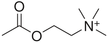
Acetylcholine
 | |
 | |
| Names | |
|---|---|
| IUPAC name
2-Acetoxy-N,N,N-trimethylethanaminium | |
| Identifiers | |
| 3DMet | B00379 |
| Abbreviations | ACh |
| ATC code | S01 |
| 1764436 | |
| 51-84-3 | |
| ChEBI | CHEBI:15355 |
| ChEMBL | ChEMBL667 |
| ChemSpider | 182 |
| DrugBank | EXPT00412 |
| EC number | 200-128-9 |
| 326108 | |
| |
| IUPHAR ligand | 294 |
| Jmol-3D images | Image |
| KEGG | C01996 |
| MeSH | Acetylcholine |
| PubChem | 187 |
| |
| UNII | N9YNS0M02X |
| Properties | |
| C 7H 16NO+ 2 | |
| Molar mass | 146.2074 g mol−1 |
| Pharmacology | |
| Elimination half-life |
2 min |
| Except where noted otherwise, data is given for materials in their standard state (at 25 °C (77 °F), 100 kPa) | |
| | |
| Infobox references | |
Acetylcholine (ACh, pron. ah-Seh-til-KO-leen) is an organic molecule that acts as a neurotransmitter in many organisms, including humans. It is an ester of acetic acid and choline, with chemical formula CH
3COO(CH
2)2N+
(CH
3)3 and systematic name 2-acetoxy-N,N,N-trimethylethanaminium.
Acetylcholine is one of many neurotransmitters in the autonomic nervous system (ANS). It acts on both the peripheral nervous system (PNS) and central nervous system (CNS) and is the only neurotransmitter used in the motor division of the somatic nervous system. Acetylcholine is also the principal neurotransmitter in all autonomic ganglia.
In cardiac tissue acetylcholine neurotransmission has an inhibitory effect, which lowers heart rate. However, acetylcholine also behaves as an excitatory neurotransmitter at neuromuscular junctions in skeletal muscle.[1]
History
Acetylcholine (ACh) was first identified in 1915 by Henry Hallett Dale for its actions on heart tissue. It was confirmed as a neurotransmitter by Otto Loewi, who initially gave it the name Vagusstoff because it was released from the vagus nerve. Both received the 1936 Nobel Prize in Physiology or Medicine for their work. Acetylcholine was also the first neurotransmitter to be identified.
Function
| Acetylcholine | |
|---|---|
| Abbreviation | ACh |
| Sources | many |
| Targets | many |
| Receptors | nicotinic; muscarinic |
| Agonists | nicotine, muscarine, succinylcholine |
| Antagonists | curare, atropine |
| Precursor | choline, acetyl-CoA |
| Synthesizing enzyme | Choline acetyltransferase (ChAT) |
| Metabolizing enzyme | Acetylcholinesterase (AChE) |
Acetylcholine has functions both in the peripheral nervous system (PNS) and in the central nervous system (CNS) as a neuromodulator. Its receptors have very high binding constants.
In the peripheral nervous system, acetylcholine activates muscles, and is a major neurotransmitter in the autonomic nervous system.
In the central nervous system, acetylcholine and the associated neurons form a neurotransmitter system, the cholinergic system, which tends to cause inhibitory actions.
In the peripheral nervous system
In the peripheral nervous system, acetylcholine activates skeletal muscles, and is a major neurotransmitter in the autonomic nervous system. Acetylcholine binds to acetylcholine receptors on skeletal muscle fibers, it opens ligand-gated sodium channels in the cell membrane. Sodium ions then enter the muscle cell, initiating a sequence of steps that finally produce muscle contraction. Although acetylcholine induces contraction of skeletal muscle, it acts via a different type of receptor (muscarinic) to inhibit contraction of cardiac muscle fibers.
In the autonomic nervous system
In the autonomic nervous system, acetylcholine is released in the following sites:
- all pre- and post-ganglionic parasympathetic neurons
- all preganglionic sympathetic neurons
- The suprarenal medullae are modified sympathetic ganglia. On stimulation by acetylcholine, the suprarenal medulla releases epinephrine and norepinephrine
- some postganglionic sympathetic fibers
- sudomotor neurons to sweat glands.
In the central nervous system
In the central nervous system, ACh has a variety of effects as a neuromodulator upon plasticity, arousal and reward. ACh has an important role in the enhancement of sensory perceptions when we wake up[2] and in sustaining attention.[3]
Damage to the cholinergic (acetylcholine-producing) system in the brain has been shown to be plausibly associated with the memory deficits associated with Alzheimer's disease.[4] ACh has also been shown to promote REM sleep.[5] Recently, it has been suggested that acetylcholine disruption may be a primary cause of depression.[6]
Pathways
There are three ACh pathways in the CNS.
- Pons to thalamus and cortex
- Magnocellular forebrain nucleus to cortex
- Septohippocampal
Structure
Acetylcholine is a polyatomic cation. It and the associated neurons form a neurotransmitter system, the cholinergic system from the brainstem and basal forebrain that projects axons to many areas of the brain. In the brainstem it originates from the Pedunculopontine nucleus and laterodorsal tegmental nucleus collectively known as the mesopontine tegmentum area or pontomesencephalotegmental complex.[7][8] In the basal forebrain, it originates from the basal optic nucleus of Meynert and medial septal nucleus:
- The pontomesencephalotegmental complex acts mainly on M1 receptors in the brainstem, deep cerebellar nuclei, pontine nuclei, locus caeruleus, raphe nucleus, lateral reticular nucleus and inferior olive.[8] It also projects to the thalamus, tectum, basal ganglia and basal forebrain.[7]
- Basal optic nucleus of Meynert acts mainly on M1 receptors in the neocortex.
- Medial septal nucleus acts mainly on M1 receptors in the hippocampus and neocortex.
In addition, ACh acts as an important "internal" transmitter in the striatum, which is part of the basal ganglia. It is released by cholinergic interneurons. In humans, non-human primates and rodents, these interneurons respond to salient environmental stimuli with stereotyped responses that are temporally aligned with the responses of dopaminergic neurons of the substantia nigra.[9][10]
Excitability and inhibition
Acetylcholine also has other effects on neurons. One effect is to cause a slow depolarization by blocking a tonically active K+
current, which increases neuronal excitability. In alternative fashion, acetylcholine can activate non-specific cation conductances to directly excite neurons.[11] An effect upon postsynaptic M4-muscarinic ACh receptors is to open inward-rectifier potassium ion channel (Kir) and cause inhibition.[12] The influence of acetylcholine on specific neuron types can be dependent upon the duration of cholinergic stimulation. For instance, transient exposure to acetylcholine (up to several seconds) can inhibit cortical pyramidal neurons via M1 type muscarinic receptors that are linked to Gq-type G-protein alpha subunits. M1 receptor activation can induce calcium-release from intracellular stores, which then activate a calcium-activated potassium conductance, which inhibits pyramidal neuron firing.[13] On the other hand, tonic M1 receptor activation is strongly excitatory. Thus, ACh acting at one type of receptor can have multiple effects on the same postsynaptic neuron, depending on the duration of receptor activation.[14] Recent experiments in behaving animals have demonstrated that cortical neurons indeed experience both transient and persistent changes in local acetylcholine levels during cue-detection behaviors.[15]
In the cerebral cortex, tonic ACh inhibits layer 4 medium spiny neurons, the main targets of thalamocortical inputs while exciting pyramidal cells in layers 2/3 and layer 5.[12] This filters out weak sensory inputs in layer 4 and amplifies inputs that reach the layers 2/3 and layer L5 excitatory microcircuits. As a result, these layer-specific effects of ACh might function to improve the signal noise ratio of cortical processing.[12] At the same time, acetylcholine acts through nicotinic receptors to excite certain groups of inhibitory interneurons in the cortex, which further dampen down cortical activity.[16]
Decision making

One well-supported function of acetylcholine (ACh) in cortex is increased responsiveness to sensory stimuli, a form of attention. Phasic increases of ACh during visual,[17] auditory[18] and somatosensory[19] stimulus presentations have been found to increase the firing rate of neurons in the corresponding primary sensory cortices. When cholinergic neurons in the basal forebrain are lesioned, animals' ability to detect visual signals was robustly and persistently impaired.[20] In that same study, animals' ability to correctly reject non-target trials was not impaired, further supporting the interpretation that phasic ACh facilitates responsiveness to stimuli. Looking at ACh's effect on thalamocortical connections, a known pathway of sensory information, in vitro application of cholinergic agonist carbachol to mouse auditory cortex enhanced thalamocortical activity.[21] In addition, Gil et al. (1997) applied a different cholinergic agonist, nicotine, and found that activity was enhanced at thalamocortical synapses.[22] This finding provides further evidence for a facilitative role of ACh in transmission of sensory information from the thalamus to selective regions of cortex.
An additional suggested function of ACh in cortex is suppression of intracortical information transmission. Gil et al. (1997) applied the cholinergic agonist muscarine to neocortical layers and found that excitatory post-synaptic potentials between intracortical synapses were depressed.[22] In vitro application of cholinergic agonist carbachol to mouse auditory cortex suppressed intracortical activity as well.[21] Optical recording with a voltage-sensitive dye in rat visual cortical slices demonstrated significant suppression in intracortical spread of excitement in the presence of ACh.[23]
Some forms of learning and plasticity in cortex appear dependent on the presence of acetylcholine. Bear et al. (1986) found that the typical synaptic remapping in striate cortex that occurs during monocular deprivation is reduced when there is a depletion of cholinergic projections to that region of cortex.[24] Kilgard et al. (1998) found that repeated stimulation of the basal forebrain, a primary source of ACh neurons, paired with presentation of a tone at a specific frequency, resulted in remapping of the auditory cortex to better suit processing of that tone.[25] Baskerville et al. (1996) investigated the role of ACh in experience-dependent plasticity by depleting cholinergic inputs to the barrel cortex of rats.[26] The cholinergic-depleted animals had a significantly reduced amount of whisker-pairing plasticity. Apart from the cortical areas, Crespo et al. (2006) found that the activation of nicotinic and muscarinic receptors in the nucleus accumbens is necessary for the acquisition of an appetitive task.[27]
ACh has been implicated in the reporting of expected uncertainty in the environment[28] based both on the suggested functions listed above and results recorded while subjects perform a behavioral cuing task. Reaction time difference between correctly cued trials and incorrectly cued trials, called the cue validity, was found to vary inversely with ACh levels in primates with pharmacologically (e.g. Witte et al., 1997) and surgically (e.g. Voytko et al., 1994) altered levels of ACh.[29][30] The result was also found in Alzheimer's disease patients (Parasuraman et al., 1992) and smokers after nicotine (an ACh agonist) consumption.[31][32] The inverse covariance is consistent with the interpretation of ACh as representing expected uncertainty in the environment, further supporting this claim.
Synthesis and degradation
Acetylcholine is synthesized in certain neurons by the enzyme choline acetyltransferase from the compounds choline and acetyl-CoA. Cholinergic neurons are capable of producing ACh. An example of a central cholinergic area is the nucleus basalis of Meynert in the basal forebrain.
The enzyme acetylcholinesterase converts acetylcholine into the inactive metabolites choline and acetate. This enzyme is abundant in the synaptic cleft, and its role in rapidly clearing free acetylcholine from the synapse is essential for proper muscle function. Certain neurotoxins work by inhibiting acetylcholinesterase, thus leading to excess acetylcholine at the neuromuscular junction, causing paralysis of the muscles needed for breathing and stopping the beating of the heart.
Receptors
There are two main classes of acetylcholine receptor (AChR), nicotinic acetylcholine receptors (nAChR) and muscarinic acetylcholine receptors (mAChR). They are named for the ligands used to activate the receptors.
Nicotinic
Nicotinic AChRs are ionotropic receptors permeable to sodium, potassium, and calcium ions. They are stimulated by nicotine and acetylcholine. They are of two main types, muscle-type and neuronal-type. The former can be selectively blocked by curare and the latter by hexamethonium. The main location of nicotinic AChRs is on muscle end plates, on autonomic ganglia (both sympathetic and parasympathetic), and in the CNS.[33]
Myasthenia gravis
The disease myasthenia gravis, characterized by muscle weakness and fatigue, occurs when the body inappropriately produces antibodies against acetylcholine nicotinic receptors, and thus inhibits proper acetylcholine signal transmission. Over time, the motor end plate is destroyed. Drugs that competitively inhibit acetylcholinesterase (e.g., neostigmine, physostigmine, or primarily pyridostigmine) are effective in treating this disorder. They allow endogenously released acetylcholine more time to interact with its respective receptor before being inactivated by acetylcholinesterase in the synaptic cleft (the space between nerve and muscle).
Muscarinic
Muscarinic receptors are metabotropic, and affect neurons over a longer time frame. They are stimulated by muscarine and acetylcholine. Muscarinic receptors are found in both the central nervous system and in the peripheral nervous system of the heart, lungs, upper GI tract, and sweat glands. ACh is sometimes used during cataract surgery to produce rapid constriction of the pupil. Atropine, occurring in the plant Deadly nightshade produces the opposite effect (anticholinergic) by blocking of the muscarinic AChRs and thereby increasing pupil size (dilation). This gives the plant both its common name (for atropine-caused heart attacks make it deadly) and its scientific name, Atropa belladonna (for women used to dilate their pupils with this plant for cosmetic purposes, "bella donna" is Italian for "beautiful lady"). It must be administered intraocularly because corneal cholinesterase metabolizes topically administered ACh before it can diffuse into the eye. Similar drugs are used to induce mydriasis (dilation of the pupil), in cardiopulmonary resuscitation and many other situations..
Drugs acting on the cholinergic system
Blocking, hindering or mimicking the action of acetylcholine has many uses in medicine. Drugs acting on the acetylcholine system are either agonists to the receptors, stimulating the system, or antagonists, inhibiting it.
| Drug | Nm | Nn | M1 | M2 | M3 |
|---|---|---|---|---|---|
| ACh, Carbachol, AChEi (Physostigmine, Galantamine, Neostigmine, Pyridostigmine) | + | + | + | + | + |
| Nicotine, Varenicline, Cotinine | + | + | |||
| Succinylcholine | +/- | ||||
| Tubocurarine, Atracurium, Cisatracurium, Rocuronium, Vecuronium, Pancuronium | - | ||||
| Epibatidine, DMPP, Decamethonium | + | ||||
| Trimethaphan, Mecamylamine, Bupropion, Dextromethorphan, Hexamethonium | - | ||||
| Muscarine, Methacholine, Oxotremorine, Bethanechol, Pilocarpine | + | + | + | ||
| Atropine, Tolterodine, Oxybutynin | - | - | - | ||
| Vedaclidine, Talsaclidine, Xanomeline, Ipratropium | - | ||||
| Pirenzepine, Telenzepine | - | ||||
| Methoctramin | - | ||||
| Darifenacin, 4-DAMP, Darifenacin, Solifenacin | - |
Nicotinic receptors are of two types: Nm and Nn. Nm is located in the neuromuscular junction which causes the contraction of skeletal muscles by way of end-plate potential (EPPs). Nn causes depolarization in autonomic ganglia resulting in post ganglionic impulse. Nicotinic receptors cause the release of catecholamine from the adrenal medulla, and also site specific excitation or inhibition in brain. Both Nm and Nn are Na + and k + channel linked but Nn is also linked with an extra Ca ++ channel.
ACh receptor agonists/antagonists
Acetylcholine receptor agonists and antagonists can either have an effect directly on the receptors or exert their effects indirectly, e.g., by affecting the enzyme acetylcholinesterase, which degrades the receptor ligand. Agonists increase the level of receptor activation, antagonists reduce it.
Associated disorders
ACh Receptor Agonists are used to treat myasthenia gravis and Alzheimer's disease.
Alzheimer's disease
Since α4β2 AchRs are reduced in Alzheimer's disease, drugs that inhibit acetylcholinesterase, e.g. galantamine hydrobromide (a competitive and reversible cholinesterase inhibitor), are commonly used in its treatment.
Direct acting
These are drugs that mimic acetylcholine on the receptor. In low doses, they stimulate the receptors, in high doses they numb them due to depolarisation block.
|
|
Cholinesterase inhibitors
Most indirect acting ACh receptor agonists work by inhibiting the enzyme acetylcholinesterase. The resulting accumulation of acetylcholine causes continuous stimulation of the muscles, glands, and central nervous system.
They are examples of enzyme inhibitors, and increase the action of acetylcholine by delaying its degradation; some have been used as nerve agents (Sarin and VX nerve gas) or pesticides (organophosphates and the carbamates). In clinical use, they are administered to reverse the action of muscle relaxants, to treat myasthenia gravis, and to treat symptoms of Alzheimer's disease (rivastigmine, which increases cholinergic activity in the brain).
Reversible
The following substances reversibly inhibit the enzyme acetylcholinesterase (which breaks down acetylcholine), thereby increasing acetylcholine levels.
- Many medications in Alzheimer's disease
- Edrophonium (differs myasthenic and cholinergic crisis)
- Neostigmine (commonly used to reverse the effect of neuromuscular blockers used in anaesthesia, or less often in myasthenia gravis)
- Physostigmine (in glaucoma and anticholinergic drug overdoses)
- Pyridostigmine (in myasthenia gravis)
- Carbamate insecticides (e.g., Aldicarb)
- Huperzine A
Irreversible
Semi-permanently inhibit the enzyme acetylcholinesterase.
- Echothiophate
- Isofluorophate
- Organophosphate Insecticides (Malathion, Parathion, Azinphos methyl, Chlorpyrifos, among others)
- Organophosphate-containing nerve agents (e.g., Sarin, VX)
Victims of organophosphate-containing nerve agents commonly die of suffocation, as they cannot relax their diaphragm.
Reactivation of acetylcholine esterase
ACh receptor antagonists
Antimuscarinic agents
- Atropine
- Ipratropium
- Scopolamine
- Tiotropium
- Diphenhydramine
Ganglionic blockers
- Mecamylamine
- Hexamethonium
- Trimethaphan
Neuromuscular blockers
|
|
|
Synthesis inhibitors
- Organic mercurial compounds, such as methylmercury, have a high affinity for sulfhydryl groups, which causes dysfunction of the enzyme choline acetyltransferase. This inhibition may lead to acetylcholine deficiency, and can have consequences on motor function.
Choline Reuptake Inhibitors
Release inhibitors
- Botulin acts by suppressing the release of acetylcholine, whereas the venom from a black widow spider (alpha-latrotoxin) has the reverse effect. ACh inhibition causes paralysis. When bitten by a black widow spider, one experiences the wastage of ACh supplies and the muscles begin to contract. If and when the supply is depleted, paralysis occurs.
Other/uncategorized/unknown
Chemical syntheses
Acetylcholine, 2-acetoxy-N,N,N-trimethylethyl ammonium chloride, is easily synthesized in a number of different ways. For example, 2-chloroethanol is reacted with trimethylamine, and the resulting N,N,N-trimethylethyl-2-ethanolamine hydrochloride, also called choline, is acetylated by acetic acid anhydride or acetylchloride, giving acetylcholine. A second method consists of reacting trimethylamine with ethylene oxide, giving N,N,N-trimethylethyl-2-ethanolamine hydroxide, which upon reaction with hydrogen chloride changes into the hydrochloride, which is further acetylated in the manner described above. Finally, acetylcholine is also formed by reacting 2-chloroethanol acetate with trimethylamine.

- A. Bayer, Ann. Chem., 142, 235 (1867).
- G. Nothnagel, Arch. Pharm., 232, 265 (1894).
- E. Fourneau, H. G. Page, Bull. Soc. Chim. France [4], 15, 544 (1914).
- H. Hopff, K. Vierling, Ger. Pat., DE 801210 (1948).
- J.K. Cline, U.S. Patent 1,957,443 (1934).
- J.K. Cline, U.S. Patent 2,012,268 (1935).
- J.K. Cline, U.S. Patent 2,013,536 (1935).
Acetylcholine is a choline molecule that has been acetylated at the oxygen atom. Because of the presence of a highly polar, charged ammonium group, acetylcholine does not penetrate lipid membranes. Because of this, when the drug is introduced externally, it remains in the extracellular space and does not pass through the blood–brain barrier. Acetylcholine does not have therapeutic value as a drug for intravenous administration because of its multi-faceted action and rapid inactivation by cholinesterase. Likewise, it is possible for a collaptoid state to develop, and arterial pressure can rapidly fall and the heart can stop. However, it is used in the form of eye drops to cause miosis during cataract surgery, which makes it advantageous because it facilitates quick post-operational recovery. A synonym of this drug is miochol.
Notes
- ↑ Campbell, N. A.; Reece, J. B. (2002). "48". Biology (6th ed.). San Francisco, CA: Pearson Education, Inc. p. 1037. ISBN 0-8053-6624-5.
- ↑ Jones, BE (2005). "From waking to sleeping: neuronal and chemical substrates". Trends in pharmacological sciences 26 (11): 578–86. doi:10.1016/j.tips.2005.09.009. PMID 16183137.
- ↑ Himmelheber, AM; Sarter, M; Bruno, JP (2000). "Increases in cortical acetylcholine release during sustained attention performance in rats". Brain research. Cognitive brain research 9 (3): 313–25. doi:10.1016/S0926-6410(00)00012-4. PMID 10808142.
- ↑ Francis PT, Palmer AM, Snape M, Wilcock GK (February 1999). "The cholinergic hypothesis of Alzheimer's disease: a review of progress". J. Neurol. Neurosurg. Psychiatr. 66 (2): 137–47. doi:10.1136/jnnp.66.2.137. PMC 1736202. PMID 10071091.
- ↑ Platt, Bettina; Riedel, Gernot (2011). "The cholinergic system, EEG and sleep". Behavioural Brain Research 221 (2): 499–504. doi:10.1016/j.bbr.2011.01.017. PMID 21238497.
- ↑ http://bbrfoundation.org/discoveries/potential-root-cause-of-depression-discovered-by-narsad-grantee
- ↑ 7.0 7.1 Woolf, NJ; Butcher, LL (1986). "Cholinergic systems in the rat brain: III. Projections from the pontomesencephalic tegmentum to the thalamus, tectum, basal ganglia, and basal forebrain". Brain Research Bulletin 16 (5): 603–37. doi:10.1016/0361-9230(86)90134-6. PMID 3742247.
- ↑ 8.0 8.1 Woolf, NJ; Butcher, LL (1989). "Cholinergic systems in the rat brain: IV. Descending projections of the pontomesencephalic tegmentum". Brain Research Bulletin 23 (6): 519–40. doi:10.1016/0361-9230(89)90197-4. PMID 2611694.
- ↑ Goldberg, J. A.; Reynolds, J. N. J. (2011). "Spontaneous firing and evoked pauses in the tonically active cholinergic interneurons of the striatum". Neuroscience 198: 27–43. doi:10.1016/j.neuroscience.2011.08.067. PMID 21925242.
- ↑ Morris, G.; Arkadir, D.; Nevet, A.; Vaadia, E.; Bergman, H. (2004). "Coincident but Distinct Messages of Midbrain Dopamine and Striatal Tonically Active Neurons". Neuron 43 (1): 133–143. doi:10.1016/j.neuron.2004.06.012. PMID 15233923.
- ↑ Haj-Dahmane, S; Andrade, R (1996). "Muscarinic activation of a voltage-dependent cation nonselective current in rat association cortex". Journal of Neuroscience 16 (12): 3848–61. PMID 8656279.
- ↑ 12.0 12.1 12.2 Eggermann, E; Feldmeyer, D (2009). "Cholinergic filtering in the recurrent excitatory microcircuit of cortical layer 4". Proceedings of the National Academy of Sciences of the United States of America 106 (28): 11753–8. doi:10.1073/pnas.0810062106. PMC 2710689. PMID 19564614.
- ↑ Gulledge, AT; Stuart, GJ (2005). "Cholinergic inhibition of neocortical pyramidal neurons". Journal of Neuroscience 25 (44): 10308–20. doi:10.1523/JNEUROSCI.2697-05.2005. PMID 16267239.
- ↑ Gulledge, AT; Bucci, DJ; Zhang, SS; Matsui, M; Yeh, HH (2009). "M1 Receptors Mediate Cholinergic Modulation of Excitability in Neocortical Pyramidal Neurons". Journal of Neuroscience 29 (31): 9888–902. doi:10.1523/JNEUROSCI.1366-09.2009. PMC 2745329. PMID 19657040.
- ↑ Parikh, V; Kozak, R; Martinez, V; Sarter, M (2007). "Prefrontal acetylcholine release controls cue detection on multiple time scales". Neuron 56 (1): 141–54. doi:10.1016/j.neuron.2007.08.025. PMC 2084212. PMID 17920021.
- ↑ Gulledge, AT; Park, SB; Kawaguchi, Y; Stuart, GJ (2007). "Heterogeneity of phasic cholinergic signaling in neocortical neurons". Journal of neurophysiology 97 (3): 2215–29. doi:10.1152/jn.00493.2006. PMID 17122323.
- ↑ Spehlmann R, Daniels JC, Smathers CC (1971). "Acetylcholine and the synaptic transmission of specific impulses to the visual cortex". Brain 94 (1): 125–38. doi:10.1093/brain/94.1.125. PMID 4324030.
- ↑ Foote SL, Freedman R, Oliver AP (March 1975). "Effects of putative neurotransmitters on neuronal activity in monkey auditory cortex". Brain Res. 86 (2): 229–42. doi:10.1016/0006-8993(75)90699-X. PMID 234774.
- ↑ Stone TW (September 1972). "Cholinergic mechanisms in the rat somatosensory cerebral cortex". J. Physiol. (Lond.) 225 (2): 485–99. PMC 1331117. PMID 5074408.
- ↑ McGaughy J, Kaiser T, Sarter M (April 1996). "Behavioral vigilance following infusions of 192 IgG-saporin into the basal forebrain: selectivity of the behavioral impairment and relation to cortical AChE-positive fiber density". Behav. Neurosci. 110 (2): 247–65. doi:10.1037/0735-7044.110.2.247. PMID 8731052.
- ↑ 21.0 21.1 Hsieh CY, Cruikshank SJ, Metherate R (October 2000). "Differential modulation of auditory thalamocortical and intracortical synaptic transmission by cholinergic agonist". Brain Res. 880 (1–2): 51–64. doi:10.1016/S0006-8993(00)02766-9. PMID 11032989.
- ↑ 22.0 22.1 Gil Z, Connors BW, Amitai Y (September 1997). "Differential regulation of neocortical synapses by neuromodulators and activity". Neuron 19 (3): 679–86. doi:10.1016/S0896-6273(00)80380-3. PMID 9331357.
- ↑ Kimura F, Fukuda M, Tsumoto T (October 1999). "Acetylcholine suppresses the spread of excitation in the visual cortex revealed by optical recording: possible differential effect depending on the source of input". Eur. J. Neurosci. 11 (10): 3597–609. doi:10.1046/j.1460-9568.1999.00779.x. PMID 10564367.
- ↑ Bear MF, Singer W (1986). "Modulation of visual cortical plasticity by acetylcholine and noradrenaline". Nature 320 (6058): 172–6. doi:10.1038/320172a0. PMID 3005879.
- ↑ Kilgard MP, Merzenich MM (March 1998). "Cortical map reorganization enabled by nucleus basalis activity". Science 279 (5357): 1714–8. doi:10.1126/science.279.5357.1714. PMID 9497289.
- ↑ Baskerville KA, Schweitzer JB, Herron P (October 1997). "Effects of cholinergic depletion on experience-dependent plasticity in the cortex of the rat". Neuroscience 80 (4): 1159–69. doi:10.1016/S0306-4522(97)00064-X. PMID 9284068.
- ↑ Crespo JA, Sturm K, Saria A, Zernig G (May 2006). "Activation of muscarinic and nicotinic acetylcholine receptors in the nucleus accumbens core is necessary for the acquisition of drug reinforcement". J. Neurosci. 26 (22): 6004–10. doi:10.1523/JNEUROSCI.4494-05.2006. PMID 16738243.
- ↑ Yu & Dayan 2005
- ↑ Witte EA, Marrocco RT (August 1997). "Alteration of brain noradrenergic activity in rhesus monkeys affects the alerting component of covert orienting". Psychopharmacology (Berl.) 132 (4): 315–23. doi:10.1007/s002130050351. PMID 9298508.
- ↑ Voytko ML, Olton DS, Richardson RT, Gorman LK, Tobin JR, Price DL (January 1994). "Basal forebrain lesions in monkeys disrupt attention but not learning and memory". J. Neurosci. 14 (1): 167–86. PMID 8283232.
- ↑ Parasuraman R, Greenwood PM, Haxby JV, Grady CL (June 1992). "Visuospatial attention in dementia of the Alzheimer type". Brain 115 (Pt 3): 711–33. doi:10.1093/brain/115.3.711. PMID 1628198.
- ↑ Witte EA, Davidson MC, Marrocco RT (August 1997). "Effects of altering brain cholinergic activity on covert orienting of attention: comparison of monkey and human performance". Psychopharmacology (Berl.) 132 (4): 324–34. doi:10.1007/s002130050352. PMID 9298509.
- ↑ Katzung, B.G. (2003). Basic and Clinical Pharmacology (9th ed.). McGraw-Hill Medical. ISBN 0-07-141092-9.
- ↑ Nałecz, Ka; Miecz, D; Berezowski, V; Cecchelli, R (Oct 2004). "Carnitine: transport and physiological functions in the brain". Molecular aspects of medicine 25 (5–6): 551–67. doi:10.1016/j.mam.2004.06.001. ISSN 0098-2997. PMID 15363641.
References
- Brenner, G.M.; Stevens, C.W. (2006). Pharmacology (2nd ed.). Philadelphia PA: W.B. Saunders. ISBN 1-4160-2984-2.
- Canadian Pharmacists Association (2000). Compendium of Pharmaceuticals and Specialties (25th ed.). Toronto ON: Webcom. ISBN 0-919115-76-4.
- Carlson, NR (2001). Physiology of Behavior (7th ed.). Needham Heights MA: Allyn and Bacon. ISBN 0-205-30840-6.
- Gershon, Michael D. (1998). The Second Brain. New York NY: HarperCollins. ISBN 0-06-018252-0.
- Siegal, A.; Sapru, H.N. (2006). "Ch. 15". Essential Neuroscience (Revised 1st ed.). Philadelphia: Lippincott, Williams & Wilkins. pp. 255–267.
- Hasselmo ME (February 1995). "Neuromodulation and cortical function: modeling the physiological basis of behavior". Behav. Brain Res. 67 (1): 1–27. doi:10.1016/0166-4328(94)00113-T. PMID 7748496. as PDF
- Yu, AJ; Dayan, P (May 2005). "Uncertainty, neuromodulation, and attention". Neuron 46 (4): 681–92. doi:10.1016/j.neuron.2005.04.026. PMID 15944135. as PDF
External links
| ||||||||||||||||||||||||||||||||||||||||