X-ray generator

An X-ray generator is a device used to generate X-rays. It is commonly used by radiographers to acquire an x-ray image of the inside of an object (as in medicine or non-destructive testing) but they are also used in sterilization or fluorescence.
Mechanism
The heart of an X-ray generator is the X-ray tube. Like any vacuum tube, the X-ray tube contains a cathode, which directs a stream of electrons into a vacuum, and an anode, which collects the electrons and is made of copper to evacuate the heat generated by the collision. When the electrons collide with the target, about 1% of the resulting energy is emitted as X-rays, with the remaining 99% released as heat. Due to the high energy of the electrons that reach relativistic speeds the target is usually made of tungsten even if other material can be used particularly in XRF applications.
A cooling system is necessary to cool the anode; many X-ray generators use water or oil recirculating systems.[2]
History
The discovery of x-rays came from experimenting with Crookes tubes, an early experimental electrical discharge tube invented by English physicist William Crookes around 1869-1875. In 1895, Wilhelm Röntgen discovered X-rays emanating from Crookes tubes and the many uses for X-rays were immediately apparent. One of the first X-ray photographs was made of the hand of Röntgen's wife. The image displayed both her wedding ring and bones. On January 18, 1896 an X-ray machine was formally displayed by H.L. Smith.
In the 1940s and 1950s, X-ray machines were used in stores to help sell footwear. These were known as fluoroscopes. However, as the harmful effects of X-ray radiation were properly considered, they finally fell out of use. Shoe-fitting use of the device was first banned by the state of Pennsylvania in 1957. (They were more a clever marketing tool to attract customers, rather than a fitting aid.)
Overview
An X-ray imaging system consists of an X-ray source or generator (X-ray tube), an image detection system which can be either a film (analog technology) or a digital capture system, and a PACS.
X-ray sources


X-ray photons are produced by an electron beam that is accelerated to a very high speed and strikes a target. The electrons that make up the beam are emitted from a heated cathode filament. The electrons are then focused and accelerated by an electrical field towards an angled anode target. The point where the electron beam strikes the target is called the focal spot. Most of the kinetic energy contained in the electron beam is converted to heat, but around 1% of the energy is converted into X-ray photons, the excess heat is dissipated via a heat sink.[3] At the focal spot, X-ray photons are emitted in all directions from the target surface, the highest intensity being around 60° to 90° from the beam due to the angle of the anode target to the approaching electron beam. There is a small round window in the X-ray tube directly above the angled target. This window allows the X-ray to exit the tube with little attenuation while maintaining a vacuum seal required for the X-ray tube operation.
X-ray machines work by applying controlled voltage and current to the X-ray tube, which results in a beam of X-rays. The beam is projected on matter. Some of the X-ray beam will pass through the object, while some is absorbed. The resulting pattern of the radiation is then ultimately detected by a detection medium including rare earth screens (which surround photographic film), semiconductor detectors, or X-ray image intensifiers.
Detection
In healthcare applications in particular, the x-ray detection system rarely consists of the detection medium. For example, a typical stationary radiographic x-ray machine also includes an ion chamber and grid. The ion chamber is basically a hollow plate located between the detection medium and the object being imaged. It determines the level of exposure by measuring the amount of x-rays that have passed through the electrically charged, gas-filled gap inside the plate. This allows for minimization of patient radiation exposure by both ensuring that an image is not underdeveloped to the point the exam needs to be repeated and ensuring that more radiation than needed is not applied. The grid is usually located between the ion chamber and object and consists of many aluminum slats stacked next to each other (resembling a polaroid lens). In this manner, the grid allows straight x-rays to pass through to the detection medium but absorbs reflected x-rays. This improves image quality by preventing scattered (non-diagnostic) x-rays from reaching the detection medium, but using a grid creates higher exam radiation doses overall.
Images taken with such devices are known as X-ray photographs or radiographs.
Applications
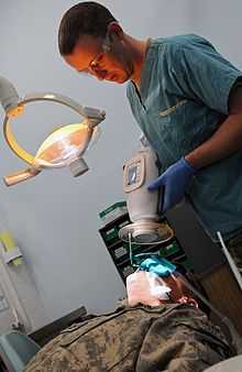
X-ray machines are used in health care for visualising bone structures and other dense tissues such as tumours. Non-medicial applications include security and material analysis.
Medicine

The two main fields in which x-ray machines are used in medicine are radiography and fluoroscopy.
Radiography is used for fast, highly penetrating images, and is usually used in areas with a high bone content. Some forms of radiography include:
- orthopantomogram — a panoramic x-ray of the jaw showing all the teeth at once
- mammography — x-rays of breast tissue
- tomography — x-ray imaging in sections
Radiotherapy — the use of x-ray radiation to treat malignant cancer cells, a non-imaging application
Fluoroscopy is used in cases where real-time visualization is necessary (and is most commonly encountered in everyday life at airport security). Some medical applications of fluoroscopy include:
- angiography — used to examine blood vessels in real time
- barium enema — a procedure used to examine problems of the colon and lower gastrointestinal tract
- barium swallow — similar to a barium enema, but used to examine the upper gastroinstestional tract
- biopsy — the removal of tissue for examination
X-rays are highly penetrating, ionizing radiation, therefore X-ray machines are used to take pictures of dense tissues such as bones and teeth. This is because bones absorb the radiation more than the less dense soft tissue. X-rays from a source pass through the body and onto a photographic cassette. Areas where radiation is absorbed show up as lighter shades of grey (closer to white). This can be used to diagnose broken or fractured bones. In fluoroscopy, imaging of the digestive tract is done with the help of a radiocontrast agent such as barium sulfate, which is opaque to X-rays.
Security

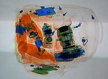
X-ray machines are used to screen objects non-invasively. Luggage at airports and student baggage at some schools are examined for possible weapons, including bombs. Prices of these Luggage X-rays vary from $70,000 to $300,000. The main parts of an X-ray Baggage Inspection System are the generator used to generate x-rays, the detector to detect radiation after passing through the baggage, signal processor unit (usually a PC) to process the incoming signal from the detector, and a conveyor system for moving baggage into the system. Portable pulsed X-ray Battery Powered X-ray Generator used in Security as shown in the figure provides EOD responders safer analysis of any possible target hazard. Prices range from $50,000 to $200,000.
Operation
When baggage is placed on the conveyor, it is moved into the machine by the operator. There is an infrared transmitter and receiver assembly to detect the baggage when it enters the tunnel. This assembly gives the signal to switch on the generator and signal processing system. The signal processing system processes incoming signals from the detector and reproduce an image based upon the type of material and material density inside the baggage. This image is then sent to the display unit.
Color classification

The colour of the image displayed depends upon the material and material density : organic material such as paper, clothes and most explosives are displayed in orange. Mixed materials such as aluminum are displayed in green. Inorganic materials such as copper are displayed in blue and non-penetrable items are displayed in black (some machines display this as a yellowish green or red). The darkness of the color depends upon the density or thickness of the material.
The material density determination is achieved by two-layer detector. The layers of the detector pixels are separated with a strip of metal. The metal absorbs soft rays, letting the shorter, more penetrating wavelengths through to the bottom layer of detectors, turning the detector to a crude two-band spectrometer. ....
Advances in X-ray technology

A film of carbon nanotubes (as a cathode) that emits electrons at room temperature when exposed to an electrical field has been fashioned into an X-ray device. An array of these emitters can be placed around a target item to be scanned and the images from each emitter can be assembled by computer software to provide a 3-dimensional image of the target in a fraction of the time it takes using a conventional X-ray device. The carbon nanotube emitters also use less energy than conventional X-ray tubes leading to lower operational costs.[4]
Engineers at the University of Missouri (MU), Columbia, have invented a compact source of x-rays and other forms of radiation. The radiation source is the size of a stick of gum and could be used to create portable x-ray scanners. A prototype handheld x-ray scanner using the source could be manufactured in as soon as three years. [5]
See also
- Fluoroscope
- Backscatter X-ray e.g., for security scanning passengers (rather than baggage)
- X-ray crystallography
- Radiography
- X-ray fluorescence
- X-ray astronomy Just detectors.
Notes
- ↑ "Deployed dentists test lightweight mobile X-ray system", Spc. Jonathan W. Thomas, 16th Mobile Public Affairs Detachment, April 21, 2011, www.army.mil. Fetched from URL on April 25, 2011.
- ↑ "X-ray Generators", NDT Resource Center. Page fetched April 21, 2011.
- ↑ "Physics of X-RAY Production".^Dead Link
- ↑ Zhang, et al. "UNC News release -- New method of using nanotube x-rays creates CT images faster than traditional scanners". Retrieved 2012-08-20.
- ↑ Editorial Staff. "MU researchers develop super compact x-ray source". Retrieved 2013-01-19.
References
- Zhang, J; Yang, G; Cheng, Y; Gao, B Qiu, Q; Lee , YZ; Lu, JP and Zhou, O (2005). "Stationary scanning X-ray source based on carbon nanotube field emitters". Applied Physics Letters 86 (May 2): 184104. Bibcode:2005ApPhL..86r4104Z. doi:10.1063/1.1923750.