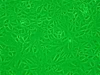
Vero cell

Vero cells are lineages of cells used in cell cultures.[1]
The Vero lineage was isolated from kidney epithelial cells extracted from an African green monkey (Chlorocebus sp.; formerly called Cercopithecus aethiops, this group of monkeys has been split into several different species). The lineage was developed on 27 March 1962, by Yasumura and Kawakita at the Chiba University in Chiba, Japan.[2] The original cell line was named "Vero" after an abbreviation of "Verda Reno", which means "green kidney" in Esperanto, while "vero" itself means "truth" also in Esperanto.[3]
Vero cells are used for many purposes, including:
- screening for the toxin of Escherichia coli, first named "Vero toxin" after this cell line, and later called "Shiga-like toxin" due to its similarity to Shiga toxin isolated from Shigella dysenteriae.
- as host cells for growing virus; for example, to measure replication in the presence or absence of a research pharmaceutical, the testing for the presence of rabies virus, or the growth of viral stocks for research purposes.
- as host cells for eukaryotic parasites, specially of the Trypanosomatids.
The Vero cell lineage is continuous and aneuploid. A continuous cell lineage can be replicated through many cycles of division and not become senescent.[4] Aneuploidy is the characteristic of having an abnormal number of chromosomes.
Vero cells are interferon-deficient; unlike normal mammalian cells, they do not secrete interferon alpha nor beta when infected by viruses.[5] However, they still have the Interferon-alpha/beta receptor so they respond normally when interferon from another source is added to the culture.
Lineages
- Vero"(ATCC No. CCL-81)
- Isolated from C. aethiops kidney on 27 Mar 1962.
- Vero 76 (ATCC No. CRL-1587)
- Isolated from Vero in 1968. It grows to a lower saturation density (cells per unit area) than the original Vero. It is useful for detecting and counting hemorrhagic fever viruses by plaque assays.
- Vero E6, also known as Vero C1008 (ATCC No. CRL-1586)
- This line is a clone from Vero 76. Vero E6 cells show some contact inhibition so are suitable for propagating viruses that replicate slowly.
- Research strains transfected with viral genes:
- Vero F6 is a cell transfected with the gene encoding HHV-1 entry protein glycoprotein-H (gH).[6] Vero F6 was transfected via a concatenated plasmid with the gH gene after a copy of the HHV-1 glycoprotein-D (gD) promoter region. In Vero lineage F6, expression of gH is under the control of the promoter region of gD. (Also F6B2; obs. F6B1.1)
See also
References
- ↑ History and Characterization of the Vero Cell Line -- A Report prepared by CDR Rebecca Sheets, Ph.D., USPHS CBER/OVRR/DVRPA/VVB for the Vaccines and Related Biological Products Advisory Committee Meeting to be held on May 12, 2000 OPEN SESSION www.fda.gov pdf
- ↑ Yasumura Y, Kawakita M (1963). "The research for the SV40 by means of tissue culture technique". Nippon Rinsho 21 (6): 1201–1219.
- ↑ Shimizu B (1993). Seno K, Koyama H, Kuroki T, ed. Manual of selected cultured cell lines for bioscience and biotechnology (in Japanese). Tokyo: Kyoritsu Shuppan. pp. 299–300. ISBN 4-320-05386-9.
- ↑ "Main Types of Cell Culture". Fundamental Techniques in Cell Culture: a Laboratory Handbook. Retrieved 2006-09-28.
- ↑ Desmyter J, Melnick JL, Rawls WE (October 1968). "Defectiveness of Interferon Production and of Rubella Virus Interference in a Line of African Green Monkey Kidney Cells (Vero)". J. Virol. 2 (10): 955–61. PMC 375423. PMID 4302013.
- ↑ Forrester A, Farrell H, Wilkinson G, Kaye J, Davis-Poynter N, Minson T (1 January 1992). "Construction and properties of a mutant of herpes simplex virus type 1 with glycoprotein H coding sequences deleted". J Virol 66 (1): 341–8. PMC 238293. PMID 1309250.
| Wikimedia Commons has media related to Vero cells. |