Vastus medialis
| Vastus medialis | |
|---|---|
| Vastus medialis | |
 | |
| Muscles of lower extremity | |
| Latin | Musculus vastus medialis or musculus vastus internus |
| Gray's | p.471 |
| Origin | Medial side of femur |
| Insertion | Quadriceps tendon |
| Artery | Femoral artery |
| Nerve | Femoral nerve |
| Actions | Extends leg |
| Dorlands /Elsevier |
Vastus medialis muscle |
The vastus medialis (/ˈvæstəs ˌmɛdiˈeɪlɨs/ or /ˈvæstəs ˌmɛdiˈælɨs/) (vastus internus or teardrop muscle) is muscle located medially in the thigh that extends the knee. The vastus medialis is part of the quadriceps muscle.
Function
The Vastus Medialis is one of five muscles that reside in the anterior compartment of the thigh. The vasti muscles appear to act largely in a co-ordinated manner throughout the control of knee extension. The vastus medialis contributes to correct tracking of the patella[1] and characteristics of the vastus medialis, including its angle of insertion, correlate with presence of patellofemoral joint pain.[2] However, this syndrome is complex and definitive evidence of causality has not yet been published.
A division of the vastus medialis muscle into two populations of fibers has been hypothesized:
- one population is thought to be long and relatively inline with the quadriceps ligament: the vastus medialis longus (VML)
- the other is thought to be shorter and more obliquely oriented with respect to the quadriceps ligament: the vastus medialis obliquus (VMO).
At the present time, there is insufficient evidence to conclusively confirm or deny this hypothesis.[3] For clinical and rehabilitation purposes, the vastus medialis is often referred to simply as the VMO in reference to its potentially important role in correct patellar tracking and prevention of patellofemoral joint syndrome.
Origin and insertion
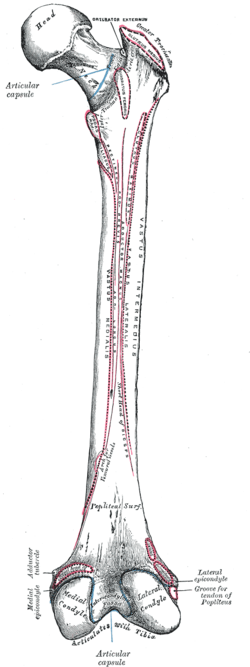
The Vastus Medialis muscle originates from a continuous line of attachment on the femur, which begins on the front and middle side (anteromedially) on the intertrochanteric line of the femur. It continues down and back (posteroinferiorly) along the pectineal line and then descends along the inner (medial) lip of the linea aspera and onto the medial supracondylar line of the femur. The fibers converge onto the inner (medial) part of the quadriceps femoris tendon and the inner (medial) border of the patella. [4]
Additional images
-
Right femur. Posterior surface.
-

Muscles of the iliac and anterior femoral regions.
-

The femoral artery.
-

Vastus medialis
-

Vastus medialis muscle
-

Vastus medialis muscle
-

Vastus medialis muscle
-

Vastus medialis muscle
-

Vastus medialis muscle
-
Muscles of thigh. Cross section.
References
- ↑ Sheehan, FT; Borotikar, BS; Behnam, AJ; Alter, KE (2012). "Alterations in in vivo knee joint kinematics following a femoral nerve branch block of the vastus medialis: Implications for patellofemoral pain syndrome". Clinical biomechanics 27 (6): 525–31. doi:10.1016/j.clinbiomech.2011.12.012. PMC 3328589. PMID 22244738.
- ↑ Jan, MH; Lin, DH; Lin, JJ; Lin, CH; Cheng, CK; Lin, YF (2009). "Differences in sonographic characteristics of the vastus medialis obliquus between patients with patellofemoral pain syndrome and healthy adults". The American journal of sports medicine 37 (9): 1743–9. doi:10.1177/0363546509333483. PMID 19521000.
- ↑ Smith, TO; Nichols, R; Harle, D; Donell, ST (2009). "Do the vastus medialis obliquus and vastus medialis longus really exist? A systematic review". Clinical anatomy 22 (2): 183–99. doi:10.1002/ca.20737. PMID 19090000.
- ↑ Drake, Richard; Vogl, Wayne; Mitchell, Adam; Gray, Henry David (2009). Gray's Anatomy for Students (2 ed.). Philadelphia, PA: Churchill Livingstone. ISBN 978-0443069529.
This article incorporates text from a public domain edition of Gray's Anatomy.
See also
- Vastus lateralis
External links
- The KNEEguru - educational site packed with knee content with sections on the quadriceps muscle
- LUC vasm
- Cross section at UV pembody/body18b
- PTCentral
- ExRx (Exercise Prescription) on the Net
- WebMD
| ||||||||||||||||||||||||||||||||||||||||||||||||||||||||||||||||||||||||