Tunica intima
| Tunica intima | |
|---|---|
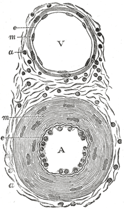 | |
| Transverse section through a small artery and vein of the mucous membrane of the epiglottis of a child. (Tunica intima is at 'e') | |
| Latin | Tunica intima |
| Gray's | subject #133 498 |
| MeSH | Tunica+Intima |
| Code | TH H3.09.02.0.01003 |
The tunica intima (or just intima) (New Latin "inner coat") is the innermost tunica (layer) of an artery or vein. It is made up of one layer of endothelial cells and is supported by an internal elastic lamina. The endothelial cells are in direct contact with the blood flow.
The tunicae of blood vessels are 3 layers: an inner, middle, and outer layer that are called, respectively, the tunica intima, the tunica media, and the tunica adventitia.
Regarding dissection, the inner coat (tunica intima) can be separated from the middle (tunica media) by a little maceration, or it may be stripped off in small pieces; but, on account of its friability, it cannot be separated as a complete membrane. It is a fine, transparent, colorless structure which is highly elastic, and, after death, is commonly corrugated into longitudinal wrinkles.
The inner coat consists of:
- A layer of pavement endothelium, the cells of which are polygonal, oval, or fusiform, and have very distinct round or oval nuclei. This endothelium is brought into view most distinctly by staining with silver nitrate.
- A subendothelial layer, consisting of delicate connective tissue with branched cells lying in the interspaces of the tissue; in arteries of less than 2 mm. in diameter the subendothelial layer consists of a single stratum of stellate cells, and the connective tissue is only largely developed in vessels of a considerable size.
- An elastic or fenestrated layer, which consists of a membrane containing a net-work of elastic fibers, having principally a longitudinal direction, and in which, under the microscope, small elongated apertures or perforations may be seen, giving it a fenestrated appearance. It was therefore called by Henle the fenestrated membrane. This membrane forms the chief thickness of the inner coat, and can be separated into several layers, some of which present the appearance of a network of longitudinal elastic fibers, and others a more membranous character, marked by pale lines having a longitudinal direction. In minute arteries the fenestrated membrane is a very thin layer; but in the larger arteries, and especially in the aorta, it has a considerable thickness.
Additional images
-

Illustration depicting structure of an artery wall
-

Vein
-

Anatomy of the arterial wall
-
Microphotography of arterial wall with calcified (violet colour) atherosclerotic plaque (haematoxillin & eosin stain)
This article incorporates text from a public domain edition of Gray's Anatomy.
External links
- Histology at OU 66_02 - "Aorta"
- Organology at UC Davis Circulatory/vessels/vessels7/vessels2 - "Bird, vessels (LM, High)"
- Tunica+intima at eMedicine Dictionary
- Image at About.com
| ||||||||||||||||||||||||||||||||