Terminal ileum
From Wikipedia, the free encyclopedia
| Terminal ileum | |
|---|---|
 | |
| Micrograph of terminal ileum with mantle cell lymphoma. H&E stain. | |
 | |
| Micrograph of terminal ileum with mantle cell lymphoma. Cyclin D1 immunostain. | |
| Latin | Pars terminalis ilei |
The terminal ileum is the most distal part of the small intestine. It connects to the cecum, the pouch between the small and the large intestine, via the ileocecal valve.
Pathology of the terminal ileum
It is of importance in medicine as it can be affected in a number of diseases,[1] including:
- Crohn's disease
- Tuberculosis
- Lymphoma
- Neuroendocrine tumours (carcinoid)
Additional images
-
Ileum
-

Small intestine
-

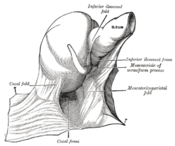
The cecal fossa. The ileum and cecum are drawn backward and upward.
References
- ↑ Cuvelier, C.; Demetter, P.; Mielants, H.; Veys, EM.; De Vos M, . (Jan 2001). "Interpretation of ileal biopsies: morphological features in normal and diseased mucosa.". Histopathology 38 (1): 1–12. doi:10.1046/j.1365-2559.2001.01070.x. PMID 11135039.
| |||||||||||||||||||||||||||||||||||||||||||||||||||
This article is issued from Wikipedia. The text is available under the Creative Commons Attribution/Share Alike; additional terms may apply for the media files.
