Supratrochlear nerve
| Nerve: Supratrochlear nerve | |
|---|---|
 | |
| Sensory areas of the head, showing the general distribution of the three divisions of the fifth nerve. (Supratrochlear nerve labeled at upper left.) | |
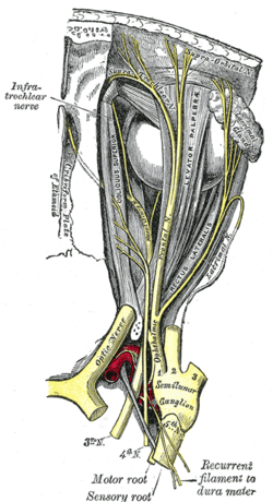 | |
| Nerves of the orbit. Seen from above. (Supratrochlear nerve visible near top.) | |
| Latin | nervus supratrochlearis |
| Gray's | p.888 |
| From | Frontal nerve |
In human cranial neuroanatomy, the supratrochlear nerve is a branch of the frontal nerve, which itself comes from the ophthalmic division of the trigeminal (or fifth) cranial nerve. It is smaller than the nearby supraorbital nerve. It passes above the pulley of the Superior oblique muscle, and gives off a descending filament that joins the infratrochlear branch of the nasociliary nerve.
The supratrochlear nerve then exits the orbit between the pulley of the superior oblique and the supraorbital foramen, curves up on to the forehead close to the bone, and ascends beneath the Corrugator supercilii and Frontalis muscles. It then divides into branches which pierce these muscles and supplies the following areas:
- skin of the lower part of the forehead, close to the midline
- conjunctiva
- skin of the upper eyelid.
Etymology
Supratrochlear means "above the trochlea". The term trochlea means "pulley" in Latin. Specifically, the trochlea referred to is a loop inside the orbit of the eye, through which the tendon of the superior oblique muscle passes.
Additional images
-

Supratrochlear nerve
-
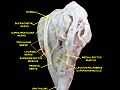
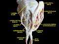
Extrinsic eye muscle. Nerves of orbita. Deep dissection.
-
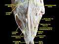
Extrinsic eye muscle. Nerves of orbita. Deep dissection.
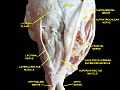
-
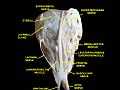
Extrinsic eye muscle. Nerves of orbita. Deep dissection.
-
Extrinsic eye muscle. Nerves of orbita. Deep dissection.
-
Extrinsic eye muscle. Nerves of orbita. Deep dissection.
See also
External links
- SUNY Figs 29:02-01
- MedEd at Loyola GrossAnatomy/h_n/cn/cn1/cnb1.htm
- lesson3 at The Anatomy Lesson by Wesley Norman (Georgetown University) (orbit2)
- cranialnerves at The Anatomy Lesson by Wesley Norman (Georgetown University) (V)
- http://www.dartmouth.edu/~humananatomy/figures/chapter_47/47-2.HTM
This article incorporates text from a public domain edition of Gray's Anatomy.
| |||||||||||||||||||||||||||||||||||||||||||||||||