Stellate reticulum
| Stellate reticulum | |
|---|---|
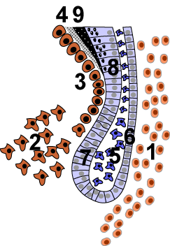 | |
| The cervical loop area: (1) dental follicle cells, (2) dental mesenchyme, (3) Odontoblasts, (4) Dentin, (5) stellate reticulum, (6) outer enamel epithelium, (7)inner enamel epithelium, (8) ameloblasts, (9) enamel. | |
| Latin | reticulum stellatum |
| Code | TE E05.04.1.1.2.3.13 |

The stellate reticulum is a group of cells located in the center of the enamel organ of a developing tooth. These cells are star shaped and synthesize glycosaminoglycans. As glycosamingoglycans are produced, water is drawn in between the cells and stretch them apart. As they are moved further away from one another, the stellate reticulum maintain contact with one another through desmosomes, resulting in their unique appearance. stellate reticulum is lost after the first layer of enamel is laid down. This brings cells in inner enamel epithelium closer to blood vessels at the periphery
References
- Orbans Oral histology and embryology – 10th ed.
Cate, A.R. Ten. Oral Histology: development, structure, and function. 5th ed. 1998. ISBN 0-8151-2952-1.
- Ross, Michael H., Gordon I. Kaye, and Wojciech Pawlina. Histology: a text and atlas. 4th edition. 2003. ISBN 0-683-30242-6.
stellate reticullum
| |||||||||||||||||||||||||||||||