Scalenus medius
| Scalenus medius | |
|---|---|
 | |
| The anterior vertebral muscles. (Scalenus medius visible at bottom center-right in red.) | |
 | |
| Muscles of the neck. Scalenus medius shown in red. | |
| Latin | Musculus scalenus medius |
| Gray's | p.396 |
| Origin | Posterior tubercles of the transverse processes of the lower six cervical vertebræ (C2, C3, C4, C5, C6 and C7) |
| Insertion | Upper surface of the first rib |
| Artery | Ascending cervical artery (branch of Inferior thyroid artery) |
| Nerve | Ventral rami of the third to eighth cervical spinal nerves |
| Actions | Elevate 1st rib, rotate the neck to the opposite side |
The Scalenus medius, also known as the middle scalene, is the largest and longest of the three scalene muscles in the human neck.
Anatomy
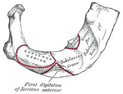
The middle scalene arises from the posterior tubercles of the transverse processes of the lower six cervical vertebræ. It descendes along the side of the vertebral column to insert by a broad attachment into the upper surface of the first rib, between the tubercle and the subclavian groove. The brachial plexus and the subclavian artery pass anterior to it. Because it elevates the upper ribs, the middle scalene muscle is also one of the accessory muscles of respiration.
Clinical Significance
It is involved in thoracic outlet syndrome, which is compression of the subclavian vessels and nerves of the brachial plexus in the region of the thoracic inlet.
Additional images
-

Position of scalenus medius (shown in red). Animation.
-

Scalenus medius (shown in red). Close up.
-

Scalenus medius (shown in red) without skull.
-

Still image.
-

Right first rib.
-

Horizontal section of the neck at about the level of the sixth cervical vertebra. Showing the arrangement of the fascia coli (scalenus medius visible at center left in green).
-
Scalenus medius muscle
-
Scalenus medius
-
Scalenus medius
-
Scalenus medius
-
Brachial plexus.Deep dissection.
See also
- Scalene muscles
- Scalenus anticus
- Scalenus posterior
- Accessory muscles of respiration
- Thoracic outlet syndrome
- Brachial plexus
- Subclavian artery
References
This article incorporates text from a public domain edition of Gray's Anatomy.
External links
| Wikimedia Commons has media related to Scalenus medius muscles. |
| ||||||||||||||||||||||||||||
.JPG)


