| Salter–Harris fractures |
|---|
|
Classification and external resources |
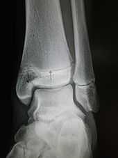
An X-ray of the left ankle showing a Salter–Harris type III fracture of medial malleolus. Black arrow demonstrates fracture line while the white arrow marks the growth plate. |
| eMedicine |
radio/613 article/1260663, orthoped/627 |
|---|
A Salter–Harris fracture is a fracture that involves the epiphyseal plate or growth plate of a bone. It is a common injury found in children, occurring in 15% of childhood long bone fractures.[1]
Types

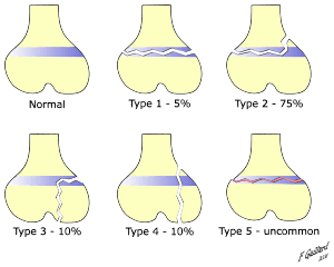
Salter Harris Fracture Types
There are nine types of Salter–Harris fractures; types I to V as described by Robert B Salter and W Robert Harris in 1963,[1] and the rarer types VI to IX which have been added subsequently:[2]
- Type I – A transverse fracture through the growth plate (also referred to as the "physis"):[3] 6% incidence
- Type II – A fracture through the growth plate and the metaphysis, sparing the epiphysis:[4] 75% incidence, takes approximately 2–3 weeks to heal.
- Type III – A fracture through growth plate and epiphysis, sparing the metaphysis:[5] 8% incidence
- Type IV – A fracture through all three elements of the bone, the growth plate, metaphysis, and epiphysis:[6] 10% incidence
- Type V – A compression fracture of the growth plate (resulting in a decrease in the perceived space between the epiphysis and diaphysis on x-ray):[7] 1% incidence
- Type VI – Injury to the peripheral portion of the physis and a resultant bony bridge formation which may produce an angular deformity (added in 1969 by Mercer Rang)[8]
- Type VII – Isolated injury of the epiphyseal plate (VII–IX added in 1982 by JA Ogden)[9]
- Type VIII – Isolated injury of the metaphysis with possible impairment of endochondral ossification
- Type IX – Injury of the periosteum which may impair intramembranous ossification
SALTER mnemonic for classification
The mnemonic "SALTR" can be used to help remember the first five types.[10][11][12] This mnemonic requires the reader to imagine the bones as long bones, with the epiphyses at the base.
- I – S = Slip (separated or straight across). Fracture of the cartilage of the physis (growth plate)
- II – A = Above. The fracture lies above the physis, or Away from the joint.
- III – L = Lower. The fracture is below the physis in the epiphysis.
- IV – TE = Through Everything. The fracture is through the metaphysis, physis, and epiphysis.
- V – R = Rammed (crushed). The physis has been crushed.
(alternatively SALTER can be used for the first 6 types - as above but adding Type V: 'E' for Everything or Epiphysis and Type VI:'R' for Ring)
Salter–Harris fracture images
| Salter–Harris fracture radiographs with insets showing fracture lines. |
|---|
| Salter–Harris I fracture of distal radius. |
| Salter–Harris II fracture of ring finger proximal phalanx. |
| Salter–Harris III fracture of big toe proximal phalanx. |
| Salter–Harris IV fracture of big toe proximal phalanx. |
|
See also
References
|
|---|
| | General | |
|---|
| | Head |
|
|---|
| | Vertebral |
|
|---|
| | Ribs | |
|---|
| | Shoulder | |
|---|
| | Arm |
|
|---|
| | Hand | |
|---|
| | Hip/femur | |
|---|
| | Leg |
|
|---|
| | Foot | |
|---|
|
|
anat (c/f/k/f, u, t/p, l)/phys/devp/cell
|
noco/cong/tumr, sysi/epon, injr
| |
|
| |
noco/cong/jaws/tumr, epon, injr
|
dent, proc (endo, orth, pros)
|
|
|
|


