Retinal pigment epithelium
| Retinal pigment epithelium | |
|---|---|
 | |
| Section of retina. (Pigmented layer labeled at bottom right.) | |
 | |
| Plan of retinal neurons. (Pigmented layer labeled at bottom right.) | |
| Latin | Stratum pigmentosum retinae, pars pigmentosa retinae |
| Gray's | subject #225 1016 |
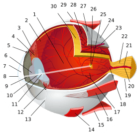
The pigmented layer of retina or retinal pigment epithelium (RPE) is the pigmented cell layer just outside the neurosensory retina that nourishes retinal visual cells, and is firmly attached to the underlying choroid and overlying retinal visual cells.[1][2]
History

The RPE was known in the 18th and 19th centuries as the pigmentum nigrum, referring to the observation that the RPE is dark (black in many animals, brown in humans); and as the tapetum nigrum, referring to the observation that in animals with a tapetum lucidum, in the region of the tapetum lucidum the RPE is not pigmented.[3]
Anatomy
The RPE is composed of a single layer of hexagonal cells that are densely packed with pigment granules.[1]
At the ora serrata, the RPE continues as a membrane passing over the ciliary body and continuing as the back surface of the iris. This generates the fibers of the dilator. Directly beneath this epithelium is the neuroepithelium (i.e., rods and cones)passes jointly with the RPE. Both, combined, are understood to be the ciliary epithelium of the embryo. The front end continuation of the retina is the posterior iris epithelium, which takes on pigment when it enters the iris[4]
When viewed from the outer surface, these cells are smooth and hexagonal in shape. When seen in section, each cell consists of an outer non-pigmented part containing a large oval nucleus and an inner pigmented portion which extends as a series of straight thread-like processes between the rods, this being especially the case when the eye is exposed to light.
Function
The RPE shields the retina from excess incoming light. It supplies omega-3 fatty acids and glucose, the former for building photoreceptive membranes, the latter for energy. Retinal is supplied by the visual vitamin A cycle. Water is removed from the retinal side to the choroid side, at a rate of 1.4-11 microliters per square centimeter per hour. It maintains balance of pH, and routinely phagocytoses the oldest outer segment discs of the photoreceptors.[5] It has a self-contained immune system, which is connected with the immune system proper to either shut down interactions when healthy, and when there is disease, it teams up with the main immune controls. Finally, it secretes substances to help build and sustain the choroid and retina.[6]
The retinal pigment epithelium also serves as the limiting transport factor that maintains the retinal environment by supplying small molecules such as amino acid, ascorbic acid and D-glucose while remaining a tight barrier to choroidal blood borne substances. Homeostasis of the ionic environment is maintained by a delicate transport exchange system.
In some clinical studies, RPE auto transplant has been used in treating Macular degeneration. Also experimental studies have reported in vitro expanded RPE used in similar studies.[7]
Pathology
In the eyes of albinos, the cells of this layer contain no pigment. Dysfunction of the RPE is found in Age-Related Macular Degeneration and Retinitis Pigmentosa.
See also
References
- ↑ 1.0 1.1 Cassin, B. and Solomon, S. (2001). Dictionary of eye terminology. Gainesville, Fla: Triad Pub. Co. ISBN 0-937404-63-2.
- ↑ Boyer MM, Poulsen GL, Nork TM. "Relative contributions of the neurosensory retina and retinal pigment epithelium to macular hypofluorescence." Arch Ophthalmol. 2000 Jan;118(1):27-31. PMID 10636410.
- ↑ Coscas, Gabriel and Felice Cardillo Piccolino (1998). Retinal Pigment Epithelium and Macular Diseases. Springer. ISBN 0-7923-5144-4.
- ↑ "eye, human."Encyclopædia Britannica from Encyclopædia Britannica 2006 Ultimate Reference Suite DVD 2009
- ↑ http://news.wustl.edu/news/Pages/25621.aspx
- ↑ http://webvision.med.utah.edu/book/part-ii-anatomy-and-physiology-of-the-retina/the-retinal-pigment-epithelium/ Webvision: The retinal pigment epithelium
- ↑ John S. et al (2013). "Choice of cell source in cell based therapies for retinal damage due to age related macular degeneration (AMD): A review". Journal of Ophthalmology.
External links
- pigment epithelium of eye at the US National Library of Medicine Medical Subject Headings (MeSH)
- BU Histology Learning System: 07902loa
- Histology at KUMC eye_ear-eye11
This article incorporates text from a public domain edition of Gray's Anatomy.
| |||||||||||||||||||||||||||||||||||||||||||||||||||||

