Posner cueing task
The Posner Cueing Task, also known as the Posner paradigm, is a neuropsychological test often used to assess attention. Formulated by Michael Posner,[1] the task assesses an individual’s ability to perform an attentional shift. It has been used and modified to assess disorders, focal brain injury, and the effects of both on spatial attention.
Method
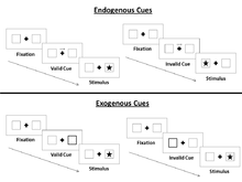
In the general paradigm, observers are seated in front of a computer screen situated at eye level. They are instructed to fixate at a central point on the screen, marked by a dot or cross. To the left and the right of the point are two boxes. For a brief period, a cue is presented on the screen. Following a brief interval after the cue is removed, a target stimulus, usually a shape, appears in either the left or right box. The observer must respond to the target immediately after detecting it. To measure reaction time (RT), a response mechanism is placed in front of the observer, usually a computer keyboard which is pressed upon detection of a target. Following a set inter-trial interval, lasting usually between 2500 and 5000 ms, the entire paradigm is repeated for a set number of trials predetermined by the experimenter.

Cues
Two major cue types are used to analyze attention based on the type of visual input. An endogenous cue is presented in the center of the screen, usually at the same location as the center of focus. It is an arrow or other directional cue pointing to the left or right box on the screen. This cue relies on input from the central visual field. An exogenous cue is presented outside of the center of focus, usually highlighting the left or right box presented on the screen. This cue relies on visual input from the peripheral visual field.
Valid and Invalid Trials
Posner devised a scheme of using valid and invalid cues across trials. In valid trials, the stimulus is presented in the area as indicated by the cue. For example, if the cue was an arrow pointing to the right, the subsequent stimulus indeed did appear in the box on the right. Conversely, in invalid trials, the stimulus is presented on the side opposite to that indicated by the cue. In this case, the arrow pointed to the right (directing attention to the right), but the stimulus in fact appeared in the box on the left. Posner used a ratio of 80% valid trials and 20% invalid trials in his original studies.[1] The observer learns that usually the cue is valid, reinforcing the tendency to direct attention to the cued side. Some trials do not present cues prior to presenting the target. These are considered neutral trials. The comparison of performance on neutral, invalid, and valid trials allows for the analysis of whether cues direct attention to a particular area and benefit or hinder attentional performance.
Overt and Covert Attention
In some studies using this paradigm, eye movements are tracked with either video-based eye tracking systems or electric potentials recorded from electrodes positioned around the eye, a process called electrooculography (EOG). This method is used to differentiate overt and covert attention. Overt attention involves directed eye movements, known as saccades, to consciously focus the eye on a target stimulus. Covert attention involves mental focus or attention to an object without significant eye movement, and is the predominant area of interest when using the Posner cueing task for research.
Experimental Findings
Variations of the Posner cueing task have been used in many studies to assess the effect of focal damage or disorders on attentional ability as well as to better understand spatial attention in healthy people. The following findings are only a few of the many results that have been established through the use of the Posner cueing task:
- Attentional shift to a target area occurs prior to any eye movement [1]
- Spatial attention is not completely reliant on conscious visual input [1]
- There are three mental operations that occur during covert orienting: disengagement of current focus, movement to selected target, and engagement of selected target [2]
- Injury to areas of the midbrain and Parkinson's disease affect orienting ability in directions in which eye movements are impaired [2][3]
- Parietal lobe damage affects the ability to orient and detect targets from invalid trials (where targets are presented on the opposite site that is directed by the cue) [2]
- Children with attention deficit hyperactivity disorder have slower reaction times in both valid and invalid trials than do typically developing children, especially to targets presented in the left visual field. Like typically developing children however, they performed better on valid trials than invalid trials.[4]
- It is mostly held that endogenous and exogenous visual spatial attention are subserved by strongly interacting, yet separable neural networks,[5][6][7][8][9] although some researchers suggest that exogenous and endogenous attention shifts are mediated by the same fronto-parietal network, consisting of the premotor cortex, posterior parietal cortex, medial frontal cortex and right inferior frontal cortex.[10]
- Cued attention is affected by age: older observers show longer engagement and delayed disengagement from cues compared to younger observers, who show increased ability in attentional shifting and disengagement relative to older observers.[11]
- Reorientation of attention to objects in a 3D space is related to proximity and unexpectedness [12]
References
- ↑ 1.0 1.1 1.2 1.3 Posner, M. I. (1980). "Orienting of attention". The Quarterly journal of experimental psychology 32 (1): 3–25. doi:10.1080/00335558008248231. PMID 7367577.
- ↑ 2.0 2.1 2.2 Posner, M. I.; Walker, J. A.; Friedrich, F. J.; Rafal, R. D. (1984). "Effects of parietal injury on covert orienting of attention". The Journal of neuroscience : the official journal of the Society for Neuroscience 4 (7): 1863–1874. PMID 6737043.
- ↑ Mari, M.; Bennett, K. M.; Scarpa, M.; Brighetti, G.; Castiello, U. (1997). "Processing efficiency of the orienting and the focusing of covert attention in relation to the level of disability in Parkinson's disease". Parkinsonism & related disorders 3 (1): 27–36. PMID 18591051.
- ↑ McDonald, S.; Bennett, K. M.; Chambers, H.; Castiello, U. (1999). "Covert orienting and focusing of attention in children with attention deficit hyperactivity disorder". Neuropsychologia 37 (3): 345–356. PMID 10199647.
- ↑ Corbetta, M., Shulman, G.L., 2002. Control of goal-directed and stimulus-driven attention in the brain. Nature Reviews Neuroscience 3, 201-215.
- ↑ Chica, A.B., Bartolomeo, P., Lupiáñez, J., 2013. Two cognitive and neural systems for endogenous and exogenous spatial attention. Behavioural Brain Research 237, 107-123.
- ↑ Hahn, B., Ross, T.J., Stein, E.A., 2006. Neuroanatomical dissociation between bottom-up and top-down processes of visuospatial selective attention. Neuroimage 32, 842-853.
- ↑ Hopfinger, J.B., West, V.M., 2006. Interactions between endogenous and exogenous attention on cortical visual processing. Neuroimage 31, 774-789.
- ↑ Kincade, J.M., Abrams, R.A., Astafiev, S.V., Shulman, G.L., Corbetta, M., 2005. An event-related functional magnetic resonance imaging study of voluntary and stimulus-driven orienting of attention. J Neurosci 25, 4593-4604.
- ↑ Peelen, M. V.; Heslenfeld, D. J.; Theeuwes, J. (2004). "Endogenous and exogenous attention shifts are mediated by the same large-scale neural network". NeuroImage 22 (2): 822–830. doi:10.1016/j.neuroimage.2004.01.044. PMID 15193611.
- ↑ Langley, L. K.; Friesen, C. K.; Saville, A. L.; Ciernia, A. T. (2011). "Timing of reflexive visuospatial orienting in young, young-old, and old-old adults". Attention, Perception, & Psychophysics 73 (5): 1546–1561. doi:10.3758/s13414-011-0108-8. PMC 3387807. PMID 21394555.
- ↑ Chen, Q.; Weidner, R.; Vossel, S.; Weiss, P. H.; Fink, G. R. (2012). "Neural Mechanisms of Attentional Reorienting in Three-Dimensional Space". Journal of Neuroscience 32 (39): 13352–13362. doi:10.1523/JNEUROSCI.1772-12.2012. PMID 23015426.
| |||||||||||||||||||||||||||||