Plantaris muscle
| Plantaris muscle | |
|---|---|
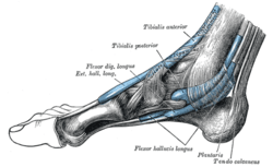 | |
| The mucous sheaths of the tendons around the ankle. Medial aspect. (Tendon of Plantaris labeled at bottom right.) | |
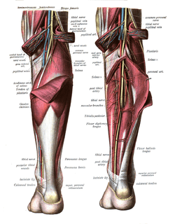 | |
| Tendon of Plantaris is labeled under the gastrocnemius (left), and folded away (right). | |
| Latin | musculus plantaris |
| Gray's | p.483 |
| Origin | Lateral supracondylar ridge of femur above lateral head of gastrocnemius |
| Insertion | Tendo calcaneus (medial side, deep to gastrocnemius tendon) |
| Artery | sural arteries |
| Nerve | tibial nerve |
| Actions | Plantar flexes foot and flexes knee |
| Antagonist | Tibialis anterior muscle |
Plantaris is one of the superficial muscles of the posterior crural compartment of the leg.
It is innervated by the tibial nerve (S1, S2) .
It is composed of a thin muscle belly and a long thin tendon. It is approximately 5-10 centimetres long, and is absent in 7-10% of the human population. It is one of the plantar flexors in the posterior compartment of the leg, along with the gastrocnemius and soleus. The plantaris is considered an unimportant muscle and mainly acts with the gastrocnemius.[1]
Structure
It arises from the inferior part of the lateral supracondylar line of the femur at a position slightly superior to the origin of the lateral head of gastrocnemius.
Also, it may arise from the oblique popliteal ligament.
Passing inferomedially posterior to the knee joint, it becomes tendinous while passing distally to insert into the tendo calcaneus, or occasionally separately inserting into the medial side of the calcaneus.
Functions
Plantaris acts to weakly:
- plantarflex the ankle joint
- flex the knee joint
Plantaris may also provide proprioceptive feedback information to the central nervous system regarding the position of the foot. The unusually high density of proprioceptive receptor end organs supports this notion.[2]
Its motor function is so minimal that its long tendon can readily be harvested for reconstruction elsewhere with little functional deficit. Often mistaken for a nerve by new medical students (and thus called the "freshman nerve"), the muscle was useful to other primates for grasping with their feet.
Clinical relevance
The plantaris is mainly used by surgeons for tendon grafts needed in other areas of the body. Although the plantaris does have little importance, there are injuries that can occur. It can be damaged in an Achilles tendon rupture. Tennis leg is a commonly known injury. It is a result of eccentric loading placed on the ankle while the knee is extended, and occurs while running or jumping. It may cause a direct trauma to the calf area. Pain and swelling are common in the injury. It is sometimes removed to treat its inflammation.[3]
Additional images
-
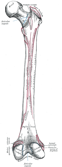
Right femur. Posterior surface.
-

Muscles of the back of the leg. Superficial layer.
-

Cross-section through middle of leg.
-

Plantaris muscle
-

MUSCLES OF LEG.POSTERIOR VIEW.
-

MUSCLES OF LEG.POSTERIOR VIEW.
References
| Wikimedia Commons has media related to Plantaris muscle. |
- ↑ DR. K. Musculoskeletal cases website: Plantaris tendon tear
- ↑ Moore, Keith L; & Dalley Arthur R (2008). Clinically Oriented Anatomy (6th ed.). Lippincott Williams and Wilkins. ISBN 978-1-60547-652-0
- ↑ http://news.bbc.co.uk/sport2/hi/football/14801756.stm
External links
- LUC plnt
- 15:st-0412 at the SUNY Downstate Medical Center
- -1274675120 at GPnotebook
- plantaris+%28muscle%29 at eMedicine Dictionary
- PTCentral
| ||||||||||||||||||||||||||||||||||||||||||||||||||||||||||||||||||||||||