Patellar ligament
| Ligament: Patellar ligament | ||
|---|---|---|
 | ||
| Right knee-joint. Anterior view. (Ligamentum patellae visible at bottom left, below patella.) | ||
| Latin | ligamentum patellae | |
| Gray's | p.340 | |
| From | patella | |
| To | tuberosity of the tibia | |
| MeSH | A02.513.514.475 | |
| Dorlands/Elsevier | l_09/12492768 | |
The patellar ligament (anterior ligament) is the central portion of the common tendon of the quadriceps femoris, which is continued from the patella to the tibial tuberosity.
Anatomy
It is a strong, flat, ligament, about 5 cm in length, which originates on the apex of the patella distally and adjoining margins of the patella and the rough depression on its posterior surface; below, it inserts on the tuberosity of the tibia; its superficial fibers are continuous over the front of the patella with those of the tendon of the Quadriceps femoris.
The medial and lateral portions of the quadriceps tendon pass down on either side of the patella, to be inserted into the upper extremity of the tibia on either side of the tuberosity; these portions merge into the capsule, as stated above, forming the medial and lateral patellar retinacula.
The posterior surface of the ligamentum patellæ is separated from the synovial membrane of the joint by a large infrapatellar pad of fat, and from the tibia by a bursa.
It is also sometimes called the "patellar tendon".[1]
Clinical significance
It can be injured in a patellar tendon rupture.
It can be used as a tissue source in the repair of other ligaments.[2]
Location of Osgood-Schlatter Disease.
See also
Additional images
-
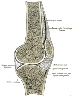
Sagittal section of right knee-joint.
-
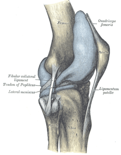
Capsule of right knee-joint (distended). Lateral aspect.
-

PATELLAR LIGAMENT.Deep dissection. Anterior view.
References
External links
- The KNEEguru - educational site packed with knee content with sections on patellar problems
- SUNY Figs 15:01-04 - "Muscles of the anterior (extensor) compartment of the leg."
- Patellar+ligament at eMedicine Dictionary
- lljoints at The Anatomy Lesson by Wesley Norman (Georgetown University) (antkneejointopenflexed)
This article incorporates text from a public domain edition of Gray's Anatomy.
| ||||||||||||||||||||||||||||||||||||||||||||||||||||||||||||||||||||||||||