Myelencephalon
| Brain: Myelencephalon | |
|---|---|
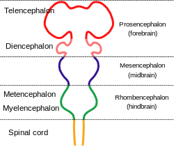 Diagram depicting the main subdivisions of the embryonic vertebrate brain. These regions will later differentiate into forebrain, midbrain and hindbrain structures | |
| Latin | Myelencephalon |
| Gray's | p.767 |
| NeuroNames | hier-695 |
| MeSH | Myelencephalon |
The myelencephalon is a subdivision of the brain used to describe the area that gives way to development of the medulla oblongata.
Development
Myelencephalon to medulla oblongata
Order of brain development in fetus:
During fetal development, divisions that give rise to the hindbrain occur at just 28 days post conception with more specific subdivisions (metencephalon, myelencephalon) taking shape at 7 weeks post conception. Final shape differentiation into the medulla oblongata can be observed at 20 weeks gestation.[1]
Medulla oblongata
Function
The medulla oblongata serves as the connection to the spinal cord from the brain, together forming the Central Nervous System. The Central Nervous system is responsible for translating and responding to peripheral stimulus. The medulla regulates vasomotor function and autonomic responses such as respiration and cardiovascular systems as well as basic reflexive activities (coughing, sneezing, swallowing, vomiting).[2]
Damage/trauma
Because of its location at the brain stem, trauma to this area can be detrimental to survival of any kind. Research shows lesions resulting from trauma can cause pulmonary edemas due to the medullas association with pulmonary function.[3] Similarly, Ischemia can also result from lesions to the medulla affecting vasomotor function.[4]
References
- ↑ Carlson, Neil R. Foundations of Behavioral Neuroscience.63-65
- ↑ Loewy, A. D., & Spyer, K. M. (Eds.). (1990). Central regulation of autonomic functions. Oxford University Press, USA.145-164
- ↑ Matsuyama, T., Okuchi, K., Nishiguchi, T., Seki, T., & Murao, Y. (2007). Neurogenic pulmonary edema caused by a medulla oblongata lesion after head trauma. The Journal of trauma, 63(3), 700.
- ↑ Kumada, M. A. M. O. R. U., Dampney, R. A., & Reis, D. J. (1979). Profound hypotension and abolition of the vasomotor component of the cerebral ischemic response produced by restricted lesions of medulla oblongata in rabbit. Relationship to the so-called tonic vasomotor center. Circulation research, 45(1), 63-70.