Lacrimal nerve
| Nerve: Lacrimal nerve | |
|---|---|
 | |
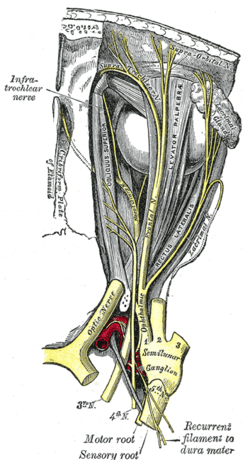
| Dissection showing origins of right ocular muscles, and nerves entering by the superior orbital fissure. | |
 | |
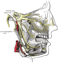
| Nerves of the orbit, and the ciliary ganglion. Side view. | |
| Latin | nervus lacrimalis |
| Gray's | p.887 |
| Innervates | lacrimal gland |
| From | ophthalmic nerve |
The lacrimal nerve is the smallest of the three branches of the ophthalmic division of the trigeminal nerve.
It sometimes receives a filament from the trochlear nerve, but this is possibly derived from the branch that goes from the ophthalmic to the trochlear nerve.
It passes forward in a separate tube of dura mater, and enters the orbit through the narrowest part of the superior orbital fissure.
In the orbit it runs along the upper border of the lateral rectus, with the lacrimal artery, and communicates with the zygomatic branch of the maxillary nerve.
It enters the lacrimal gland and gives off several filaments, which supply sensory innervation to the gland and the conjunctiva.
Then, it pierces the orbital septum, and ends in the skin of the upper eyelid, joining with filaments of the facial nerve.
The lacrimal nerve is occasionally absent, and its place is then taken by the zygomaticotemporal branch of the maxillary nerve. Sometimes the latter branch is absent, and a continuation of the lacrimal nerve is substituted for it.
Functions
It provides sensory innervations for the lacrimal gland, conjunctiva, and the lateral upper eyelids.
The zygomatic nerve carries sensory fibers from the skin and mucous membranes. It also carries post-synaptic parasympathetic fibers (originating in the pterygopalatine ganglion) to the lacrimal nerve via a communication. These fibers will eventually provide innervation to the lacrimal gland.
Additional images
-

Nerves of the orbit. Seen from above.
-

Distribution of the maxillary and mandibular nerves, and the submaxillary ganglion.
-

Sensory areas of the head, showing the general distribution of the three divisions of the fifth nerve.
-

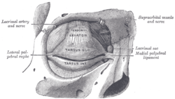
The tarsi and their ligaments. Right eye; front view.
-
Extrinsic eye muscle. Nerves of orbita. Deep dissection.
-
Extrinsic eye muscle. Nerves of orbita. Deep dissection.
-
Extrinsic eye muscle. Nerves of orbita. Deep dissection.
-
Extrinsic eye muscle. Nerves of orbita. Deep dissection.
-
Extrinsic eye muscle. Nerves of orbita. Deep dissection.
-
Extrinsic eye muscle. Nerves of orbita. Deep dissection.
-
Extrinsic eye muscle. Nerves of orbita. Deep dissection.
-
Extrinsic eye muscle. Nerves of orbita. Deep dissection.
External links
- 29:16-0102 at the SUNY Downstate Medical Center - "Orbits and Eye: The Lacrimal Gland"
- n2a4p2 at the University of Arkansas for Medical Sciences - "Branches of Trigeminal Nerve, Lateral View"
- MedEd at Loyola GrossAnatomy/h_n/cn/cn1/cnb1.htm
- cranialnerves at The Anatomy Lesson by Wesley Norman (Georgetown University) (VII)
This article incorporates text from a public domain edition of Gray's Anatomy.
| |||||||||||||||||||||||||||||||||||||||||||||||||


