"Kneecap" redirects here. For other uses, see
NECAP.
The patella (//), also known as the kneecap or kneepan, is a thick, circular-triangular bone which articulates with the femur (thigh bone) and covers and protects the anterior articular surface of the knee joint. The patella appears in the majority of vertebrates with a femur, for example mice, cats, and birds, but not whales or most reptiles and amphibians such as snakes or frogs.
In humans, the patella is the largest sesamoid bone in the body. Babies are born with a patella of soft cartilage which begins to ossify into bone at about three years of age.
Structure

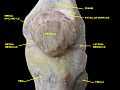
Human left patella - anterior aspect
The patella is roughly triangular in shape with its base facing proximally (towards the torso) and its tip (apex patellae) facing distally (towards the feet). Its anterior and posterior surfaces are joined laterally (left/right) by a thinner margin and medially (towards centre) by a thicker margin.[1]
Anterior surface
The anterior surface can be divided into three parts:[1]
- The upper third is coarse, flattened, and rough; it serves for the attachment of the tendon of the quadriceps and often has exostoses.
- The middle third has numerous vascular canaliculi.
- The lower third includes the distal apex which serves as the origin of the patellar ligament.
Posterior surface

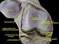
Human left patella - posterior aspect
The posterior surface is divided into two parts.[1]
The upper three-quarters articulates with the femur and is subdivided into a medial and a lateral facet by a vertical ledge which varies in shape. Four main types of articular surface can be distinguished:
- Most commonly the medial articular surface is smaller than the lateral.
- Sometimes both articular surfaces are virtually equal in size.
- Occasionally, the medial surface is hypoplastic or
- the central ledge is only indicated.
In the adult the articular surface is about 12 cm2 (1.9 sq in) and covered by cartilage, which can reach a maximal thickness of 6 mm (0.24 in) in the centre at about 30 years of age.
The lower part of the posterior surface has vascular canaliculi filled and is filled by fatty tissue, the infrapatellar fat pad.
Muscles
It is attached to the tendon of the quadriceps femoris muscle, which contracts to extend/straighten the knee. The vastus intermedius muscle is attached to the base of patella. The vastus lateralis and vastus medialis are attached to lateral and medial borders of patella respectively.
The patella is stabilized by the insertion of vastus medialis and the prominence of the anterior femoral condyles, which prevent lateral dislocation during flexion. The retinacular fibres of the patella also stabilize it during exercise.
Variation
Emarginations (i.e. patella emarginata, a "missing piece") are common laterally on the proximal edge.[1] Bipartite patellas are the result of an ossification of a second cartilaginous layer at the location of an emargination. Previously, bipartite patellas were explained as the failure of several ossification centres to fuse, but this idea has been rejected. Partite patellas occur almost exclusively in men. Tripartite and even multipartite patellas occur.
Development
In the patella an ossification centre develops between the ages 3–6 years.[1]
In animals
The patella has convergently evolved in placental mammals and birds; most marsupials have only rudimentary, non-ossified patellae although a few species possess a bony patella.[2] A patella is also present in the living monotremes, the platypus and the echidna. In more primitive tetrapods, including living amphibians and most reptiles (except some Lepidosaurs), the muscle tendons from the upper leg are attached directly to the tibia, and a patella is not present.[3]
Function
The primary functional role of the patella is knee extension. The patella increases the leverage that the tendon can exert on the femur by increasing the angle at which it acts.
Clinical relevance
Dislocation
Patellar dislocations occur with significant regularity, particular in young female athletes.[4] It involves the patella sliding out of its position on the knee, most often laterally, and may be associated with extremely intense pain and swelling.[5] The patella can be tracked back into the groove with an extension of the leg, and therefore sometimes returns into the proper position on its own.[5]
History
Etymology
The etymology of patella presents an interesting case of convergent etymology. In the Western world, 'patella' originated in the late 17th century from the diminutive form of Latin patina, meaning shallow dish.[6] The use of this term in Eastern medicine, however, predates the Western etymology. As early as the 13th century, traditional Ayurveda practitioners in the Indian subcontinent referred to the kneecap as paṭēla haḍḍī (Hindi: पटेल हड्डी), literally 'farmer's bone' because the grueling life of a patidar - and Indian land tenant, commonly resulted in injuries and inflammation of the joints, particularly the knee. The term was subsequently shortened to just 'patela'. During the British Raj period, Western medicine was introduced to India, and the two etymologies converged into the modern medical use, with two 'L's.
See also
This article uses anatomical terminology; for an overview, see anatomical terminology.
Additional images
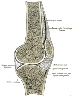
| Right knee-joint. Anterior view. |
| Sagittal section of right knee-joint. |
| Capsule of right knee-joint (distended). Lateral aspect. |
| Front and medial aspect of right thigh. |
| Lateral aspect of right leg. |
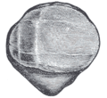
| Posterior surface of right patella |
| Patella - anterior surface |
| Patella - posterior surface. |
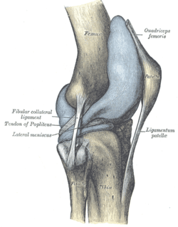
| Knee joint.Deep dissection. Anteromedial view. |
| Knee joint. Deep dissection. Anterior view. |
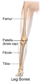
| Location of Kneecap relative to Knee Anatomy |
|
References
- ↑ 1.0 1.1 1.2 1.3 1.4 Platzer, Werner (2004). Color Atlas of Human Anatomy, Vol. 1: Locomotor System (5th ed.). Thieme. p. 194. ISBN 3-13-533305-1.
- ↑ Herzmark MH (1938). "The Evolution of the Knee Joint". J Bone Joint Surg Am 20 (1): 77–84. Retrieved 2007-11-17.
- ↑ Romer, Alfred Sherwood; Parsons, Thomas S. (1977). The Vertebrate Body. Philadelphia, PA: Holt-Saunders International. p. 205. ISBN 0-03-910284-X.
- ↑ Palmu, S.; Kallio, P.E.; Donell, S.T.; Helenius, I.; Nietosvaara, Y. (2008). "Acute patellar dislocation in children and adolescents: A randomized clinical trial". Journal of Bone and Joint Surgery 90 (3): 463–470. doi:10.2106/JBJS.G.00072. PMID 18310694.
- ↑ 5.0 5.1 Dath, R.; Chakravarthy, J.; Porter, K.M. (2006). "Patella Dislocations". Trauma 8: 5–11. doi:10.1191/1460408606ta353ra.
- ↑ http://www.etymonline.com/index.php?term=patella
External links
|
|---|
| | Femur |
|
|---|
| | Crus |
|
|---|
| | Foot |
| |
|---|
| | |
|---|
| Other |
phalanges of the foot |
|---|
|
|---|
|
|
anat (c/f/k/f, u, t/p, l)/phys/devp/cell
|
noco/cong/tumr, sysi/epon, injr
| |
|
|
|