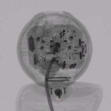
Industrial computed tomography

Industrial computed tomography (CT) scanning is any computer-aided tomographic process, usually x-ray computed tomography, that (like its medical imaging counterparts) uses irradiation (usually with x-rays) to produce three-dimensional representations of the scanned object both externally and internally. Industrial CT scanning has been used in many areas of industry for internal inspection of components. Some of the key uses for CT scanning have been flaw detection, failure analysis, metrology, assembly analysis and reverse engineering applications.[1][2] Just as in medical imaging, industrial imaging includes both nontomographic radiography (industrial radiography) and computed tomographic radiography (computed tomography).
Classification
| Type | Power range | Resolution |
|---|---|---|
| Nano | n/a | less than 1 µm |
| Low power | 0–110 kV | greater than 1 µm |
| Mid-power | 110kV–999 kV | greater than 1 µm |
| High power | greater than 1 MV | greater than 1 µm |
Types of scanners

Fan/line beam scanners-translate[3][4]
Line scanners are the first generation of industrial CT Scanners. X-rays are produced and the beam is collimated to create a line. The X-ray line beam is then translated across the part and data is collected by the detector. The data is then reconstructed to create a 3-D Volume rendering of the part.
Cone beam scanners-rotate[4]
During the CT scan the part is placed on a rotary table. As the part rotates the cone of X-rays produce about 1300 2D images which are collected by the detector. The 2D images are then processed to create a 3D volume rendering of the external and internal geometries of the part.

History
Industrial CT scanning technology was introduced in 1972 with the invention of the CT scanner by G. Hounsfield. The invention earned him a Nobel Prize in medicine, which he shared with Allan Cormack.[5][6]
Many advances in CT scanning have allowed for its use in the industrial field for metrology in addition to the visual inspection primarily used in the medical field (medical CT scan).
Analysis/inspection techniques
Various inspection techniques include:[7]
Part to CAD comparisons, part to part comparisons, assembly / defect analysis, void analysis, wall thickness analysis, and generation of CAD data for reverse engineering requirements and GD&T (geometric dimensioning and tolerance) analysis to meet PPAP (production part approval process) requirements.
Assembly
One of the most recognized forms of analysis using CT is assembly or visual analysis. CT scanning has been largely used for medical purposes as an imaging tool to supplement medical ultrasonography and X-rays as well as for screening for disease and preventative medicine. For industrial CT scanning, the ability to see inside a component is beneficial since internal components can be seen in their functioning position. Also, devices can be analyzed without disassembly. Some software programs for industrial CT scanning allow for measurements to be taken from the CT dataset volume rendering. These measurements are useful for determining the clearances between assembled parts or simply a dimension of an individual feature.
Part comparisons (part to part or part to CAD)
In today’s market parts can be manufactured around the world: designed in one country, machined in another and assembled in a third. Verification of the part to the original CAD design is critical, especially if the part is to be used in an assembly. Industrial computed tomography allows for a comparison of parts to one another or parts to CAD data. The deviations for both external and internal geometries can be shown on the surface colour map chromatically on the 3D representation or by whisker plots in the 2D windows. This process is beneficial when comparing the same part from various suppliers, studying the differences in parts from one cavity to another cavity from the same mould, or verifying the design to the part.
Void, crack and defect detection

Traditionally, determining defects, voids and cracks within an object would require destructive testing. CT scanning can detect internal features and flaws displaying this information in 3D without destroying the part. Industrial CT scanning (3D X-ray) is used to detect flaws inside a part such as porosity,[8] an inclusion, or a crack.[9] In some software programs the porosity within a part is categorized by colour based on their respective sizes.
Metal casting and moulded plastic components are typically prone to porosity because of cooling processes, transitions between thick and thin walls, and material properties. Void analysis can be used to locate, measure, and analyze voids inside plastic or metal components.
Generation of CAD data for reverse engineering requirements
A CAD file can be generated from the CT data set, which is particularly useful in reverse engineering applications and product development. Exported CAD file formats are recognized by many software such as CAD, FEA, and CFD. The CAD file created by CT scanning can not only show the external components, but the internal as well. This allows for first-time rapid prototyping of internal components without the task of creating an entirely new CAD file by hand.
Geometric dimensioning and tolerancing analysis
Traditionally, without destructive testing, full metrology has only been performed on the exterior dimensions of components. If a highly detailed component requires inspection, the conventional method of inspection would be to fixture the part to create specified datum reference plane and go through a timely CMM touch-probe inspection process or use a vision system to map exterior surfaces. Past internal inspection methods would require using a 2D X-ray of the component or the use of destructive testing. Industrial CT scanning allows for full metrology of the CT datasets allowing for an analysis of GD&T points to meet the PPAP requirement.
See also
References
- ↑ Flisch, A., et al. Industrial Computer Tomography in Reverse Engineering Applications. DGZfP-Proceedings BB 67-CD Paper 8, Computerized Tomography for Industrial Applications and Image Processing in Radiology, March 15–17, 1999, Berlin, Germany.
- ↑ Woods, Susan. "3-D CT inspection offers a full view of microparts", Nov. 1, 2010. http://micromanufacturing.com/showthread.php?t=876
- ↑ "CT Services – Overview." Jesse Garant & Associates. August 17, 2010. http://www.jgarantmc.com/ct-services.html
- ↑ 4.0 4.1 Hofmann, J., Flisch, A., Obrist, A., Adaptive CT scanning-mesh based optimisation methods for industrial X-ray computer tomography applications. NDT&E International (37), 2004, pp. 271–278.
- ↑ Zoofan, Bahman. "3D Micro-Tomography – A Powerful Engineering Tool". 3D Scanning Technologies. July 5, 2010 <http://www.3dscanningtechnologies.com/ComputedTomographyPage.php?3D_Micro-Tomography_-_A_Powerful_Engineering_Tool-4/>.
- ↑ Noel, Julien. "Advantages of CT in 3D Scanning of Industrial Parts. August 18, 2010. <http://www.3dscanningtechnologies.com/pdfs/parts.pdf>
- ↑ "Reducing Preproduction Inspection Costs with Industrial (CT) Computed Tomography." Commercial Micro Manufacturing Magazine for the global micro manufacturing technology industry, August 2010. http://www.micromanu.com/x/guideArchiveArticle.html?id=1695
- ↑ Lambert, J.; Chambers, A. R.; Sinclair, I.; Spearing, S. M. (2012). "3D damage characterisation and the role of voids in the fatigue of wind turbine blade materials". Composites Science and Technology 72 (2): 337. doi:10.1016/j.compscitech.2011.11.023.
- ↑ Bull, D. J.; Helfen, L.; Sinclair, I.; Spearing, S. M.; Baumbach, T. (2013). "A comparison of multi-scale 3D X-ray tomographic inspection techniques for assessing carbon fibre composite impact damage". Composites Science and Technology 75: 55. doi:10.1016/j.compscitech.2012.12.006.