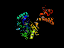
Hydrogenase
A hydrogenase is an enzyme that catalyses the reversible oxidation of molecular hydrogen (H2), as shown in below:
- (1) H2 + Aox → 2H+ + Ared
- (2) 2H+ + Dred → H2 + Dox
Hydrogen uptake (1) is coupled to the reduction of electron acceptors such as oxygen, nitrate, sulfate, carbon dioxide, and fumarate. On the other hand, proton reduction (2) is coupled to the oxidation of electron donors such as ferredoxin (FNR), and serves to dispose excess electrons in cells (essential in pyruvate fermentation). Both low-molecular weight compounds and proteins such as FNRs, cytochrome c3, and cytochrome c6 can act as physiological electron donors or acceptors for hydrogenases.[1]
Structural classification

Hydrogenases are classified as one of the following three types based on metal atoms composing the active site: [NiFe], [FeFe], and [Fe]-only. Until 2004, the [Fe]-only hydrogenase was believed to be "metal-free". Then, Thauer et al. showed that the metal-free hydrogenases in fact contain iron atom in its active site. As a result, those enzymes previously classified as "metal-free" are now named [Fe]-only hydrogenases. This protein contains only a mononuclear Fe active site and no iron-sulfur clusters, in contrast to the [FeFe] hydrogenases. [NiFe] and [FeFe] hydrogenases have some common features in their structures: Each enzyme has an active site and a few Fe-S clusters that are buried in protein. The active site, which is believed to be the place where catalysis takes place, is also a metallocluster, and each metal is coordinated by carbon monoxide (CO) and cyanide (CN-) ligands.[2]
[NiFe] hydrogenase

The [NiFe] hydrogenases are heterodimeric proteins consisting of small (S) and large (L) subunits. The small subunit contains three iron-sulfur clusters while the large subunit contains the active site, a nickel-iron centre which is connected to the solvent by a molecular tunnel.[3] In some [NiFe] hydrogenases, one of the Ni-bound cysteine residues is replaced by selenocysteine. On the basis of sequence similarity, however, the [NiFe] and [NiFeSe] hydrogenases should be considered a single superfamily. To date, periplasmic, cytoplasmic, and cytoplasmic membrane-bound hydrogenases have been found. The [NiFe] hydrogenases, when isolated, are found to catalyse both H2 evolution and uptake, with low-potential multihaem cytochromes such as cytochrome c3 acting as either electron donors or acceptors, depending on their oxidation state. Generally speaking, however, [NiFe] hydrogenases are more active in oxidizing H2.
Like [FeFe] hydrogenases, [NiFe] hydrogenases are known to be deactivated by molecular oxygen (O2). Recently, a novel hydrogenase from Ralstonia eutropha have been found to be oxygen-tolerant.[4] This finding increased hope that hydrogenases can be used in photosynthetic production of molecular hydrogen via splitting water.
[FeFe] hydrogenase

The hydrogenases containing a di-iron center with a bridging dithiolate cofactor are called [FeFe] hydrogenases.[5] Three families of [FeFe] hydrogenases are recognized:
- cytoplasmic, soluble, monomeric hydrogenases, found in strict anaerobes such as Clostridium pasteurianum and Megasphaera elsdenii. They are extremely sensitive to inactivation by dioxygen (O2) and catalyse both H2 evolution and uptake.
- periplasmic, heterodimeric hydrogenases from Desulfovibrio spp., which can be purified aerobically and catalyse mainly H2 oxidation.
- soluble, monomeric hydrogenases, found in chloroplasts of green alga Scenedesmus obliquus, catalyses H2 evolution. The [Fe2S2] ferredoxin functions as natural electron donor linking the enzyme to the photosynthetic electron transport chain.
In contrast to [NiFe] hydrogenases, [FeFe] hydrogenases are generally more active in production of molecular hydrogen. Turnover frequency (TOF) in the order of 10,000 s-1 have been reported in literature for [FeFe] hydrogenases from clostridium pasteurianum.[6] This has led to intense research focusing on use of [FeFe] hydrogenase for sustainable production of H2.[7]
[Fe]-only hydrogenase

5,10-methenyltetrahydromethanopterin hydrogenase (EC 1.12.98.2) found in methanogenic Archaea contains neither nickel nor iron-sulfur clusters but an iron-containing cofactor that was recently characterized by X-ray diffraction.[8]
Unlike the other two types, [Fe]-only hydrogenases are found only in some hydrogenotrophic methanogenic archaea. They also feature a fundamentally different enzymatic mechanism in terms of redox partners and how electrons are delivered to the active site. In [NiFe] and [FeFe] hydrogenases, electrons travel through a series of metallorganic clusters that comprise a long distance; the active site structures remain unchanged during the whole process. In [Fe]-only hydrogenases, however, electrons are directly delivered to the active site via a short distance. Methenyl-H4MPT+, a cofactor, directly accepts the hydride from H2 in the process. [Fe]-only hydrogenase is also known as H2-forming methylenetetrahydromethanopterin (methylene-H4MPT) dehydrogenase, because its function is the reversible reduction of methenyl-H4MPT+ to methylene-H4MPT.[9] The hydrogenation of a methenyl-H4MPT+ occurs instead of H2 oxidation/production, which is the case for the other two types of hydrogenases. While the exact mechanism of the catalysis is still under study, recent finding suggests that molecular hydrogen is first heterolytically cleaved by Fe(II), followed by transfer of hydride to the carbocation of the acceptor.[10]
Mechanism
The molecular mechanism by which protons are converted into hydrogen molecules within hydrogenases is still under extensive study. One popular approach employs mutagenesis to elucidate roles of amino acids and/or ligands in different steps of catalysis such as intramolecular transport of substrates. For instance, Cornish et al. conducted mutagenesis studies and found out that four amino acids located along the putative channel connecting the active site and protein surface are critical to enzymatic function of [FeFe] hydrogenase from clostridium pasteurianum (CpI). [11] On the other hand, one can also rely on computational analysis and simulations. Nilsson Lill and Siegbahn have recently taken this approach in investigating the mechanism by which [NiFe] hydrogenases catalyze H2 cleavage.[12] The two approaches are complementary and can benefit one another. In fact, Cao and Hall combined both approaches in developing the model that describes how hydrogen molecules are oxidized or produced within the active site of [FeFe] hydrogenases. [13] While more research and experimental data are required to complete our understanding of the mechanism, these findings have allowed scientists to apply the knowledge in, e.g., building artificial catalysts mimicking active sites of hydrogenases. [14]
Biological function
Assuming that the Earth's atmosphere was initially rich in hydrogen, scientists hypothesize that hydrogenases were evolved to generate energy from/as molecular H2. Accordingly, hydrogenases can either help microorganisms to proliferate under such conditions, or to set up ecosystems empowered by H2.[15]Microbian communities driven by molecular hydrogen have, in fact, been found in deep-sea settings where other sources of energy from photosynthesis are not available. Based on these grounds, the primary role of hydrogenases are believed to be energy generation, either for itself or others within the community.
Recent studies have revealed other biological functions of hydrogenases. To begin with, bidirectional hydrogenases can also act as "valves" to control excess reducing equivalents, especially in photosynthetic microorganisms. Such a role makes hydrogenases play a vital role in anaerobic metabolism.[16][17] Moreover, hydrogenases may also be involved in membrane-linked energy conservation through the generation of a transmembrane protonmotive force.[15]There is a possibility that hydrogenases have been responsible for bioremediation of chlorinated compounds. Hydrogenases proficient in H2 uptake can help heavy metal contaminants to be recovered in intoxicated forms. Interestingly, these uptake hydrogenases have been recently discovered in pathogenic bacteria and parasites and are believed to be involved in their virulence[15].
Applications
Hydrogenases were first discovered in the 1930s,[18] and they have since attracted interest from many researchers including inorganic chemists who have synthesized a variety of hydrogenase mimics. Understanding the catalytic mechanism of hydrogenase might help scientists design clean biological energy sources, such as algae, that produce hydrogen.[19]
Biological hydrogen production
Various systems are capable of splitting water into O2 and H+ from incident sunlight. Likewise, numerous catalysts, either chemical or biological, can reduce the produced H+ into H2. Different catalysts require unequal overpotential for this reduction reaction to take place. Hydrogenases are attractive since they require a relatively low overpotential. In fact, its catalytic activity is more effective than platinum, which is the best known catalyst for H2 evolution reaction.[20] Among three different types of hydrogenases, [FeFe] hydrogenases is considered as a strong candidate for an integral part of the solar H2 production system since they offer an additional advantage of high TOF (over 9000 s-1)[6].
Low overpotential and high catalytic activity of [FeFe] hydrogenases are accompanied by high O2 sensitivity. It is necessary to engineer them O2-tolerant for use in solar H2 production since O2 is a by-product of water splitting reaction. Past research efforts by various groups around the world have focused on understanding the mechanisms involved in O2-inactivation of hydrogenases.[21][22] For instance, Stripp et al. relied on protein film electrochemistry and discovered that O2 first converts into a reactive species at the active site of [FeFe] hydrogenases, and then damages its [4Fe-4S] domain.[23] Cohen et al. investigated how oxygen can reach the active site that is buried inside the protein body by molecular dynamics simulation approach; their results indicate that O2 diffuses through mainly two pathways that are formed by enlargement of and interconnection between cavities during dynamic motion.[24] These works, in combination with other reports, suggest that inactivation is governed by two phenomena: diffusion of O2 to the active site, and destructive modification of the active site.
Despite these findings, research is still under progress for engineering oxygen tolerance in hydrogenases. While researchers have found oxygen-tolerant [NiFe] hydrogenases, they are only efficient in hydrogen uptake and not production[21]. Bingham et al.’s recent success in engineering [FeFe] hydrogenase from clostridium pasteurianum was also limited to retained activity (during exposure to oxygen) for H2 consumption, only.[25]
Biochemical classification
EC 1.2.1.2 hydrogen dehydrogenase (hydrogen:NAD+ oxidoreductase)
- H2 + NAD+ = H+ + NADH
EC 1.12.1.3 hydrogen dehydrogenase (NADP) (hydrogen:NADPH+ oxidoreductase)
- H2 + NADP+ = H+ + NADPH
EC 1.12.2.1 cytochrome-c3 hydrogenase (hydrogen:ferricytochrome-c3 oxidoreductase)
- 2H2 + ferricytochrome c3 = 4H+ + ferrocytochrome c3
EC 1.12.7.2 ferredoxin hydrogenase (hydrogen:ferredoxin oxidoreductase)
- H2 + oxidized ferredoxin = 2H+ + reduced ferredoxin
EC 1.12.98.1 coenzyme F420 hydrogenase (hydrogen:coenzyme F420 oxidoreductase)
- H2 + coenzyme F420 = reduced coenzyme F420
EC 1.12.99.6 hydrogenase (acceptor) (hydrogen:acceptor oxidoreductase)
- H2 + A = AH2
EC 1.12.5.1 hydrogen:quinone oxidoreductase
- H2 + menaquinone = menaquinol
EC 1.12.98.2 5,10-methenyltetrahydromethanopterin hydrogenase (hydrogen:5,10-methenyltetrahydromethanopterin oxidoreductase)
- H2 + 5,10-methenyltetrahydromethanopterin = H+ + 5,10-methylenetetrahydromethanopterin
EC 1.12.98.3 Methanosarcina-phenazine hydrogenase [hydrogen:2-(2,3-dihydropentaprenyloxy)phenazine oxidoreductase]
- H2 + 2-(2,3-dihydropentaprenyloxy)phenazine = 2-dihydropentaprenyloxyphenazine
References
- ↑ Vignais, P.M., Billoud, B. and Meyer, J. (2001). "Classification and phylogeny of hydrogenases". FEMS Microbiol. Rev. 25 (4): 455–501. PMID 11524134.
- ↑ Fontecilla-Camps, J.C., Volbeda, A., Cavazza, C., Nicolet Y. (2007). "Structure/function relationships of [NiFe]- and [FeFe]-hydrogenases". Chem Rev 107 (10): 4273–4303. doi:10.1021/cr050195z. PMID 17850165.
- ↑ Liebgott PP, Leroux F, Burlat B, Dementin S, Baffert C, Lautier T, Fourmond V, Ceccaldi P, Cavazza C, Meynial-Salles I, Soucaille P, Fontecilla-Camps JC, Guigliarelli B, Bertrand P, Rousset M, Léger C. (2010). "Relating diffusion along the substrate tunnel and oxygen sensitivity in hydrogenase". Nat. Chem. Biol. 6 (1): 63–70. doi:10.1038/nchembio.276. PMID 19966788.
- ↑ Burgdorf, T., Buhrke, T., van der Linden, E., Jones, A., Albracht, S. and Friedrich, B. (2005). "[NiFe]-Hydrogenases of Ralstonia eutropha H16: Modular Enzymes for Oxygen-Tolerant Biological Hydrogen Oxidation". J. Mol. Microbiol. Biotechnol. 10: 181–196. doi:10.1159/000091564. PMID 16645314.
- ↑ Berggren, G., Adamska, A., Lambertz, C., Simmons, T. R., Esselborn, J., Atta, A., Gambarelli, S., Mouesca, J.-M., Reijerse, E., Lubitz, W., Happe, T., Artero, V., Fontecave, M. (2013). "Biomimetic assembly and activiation of [FeFe]-hydrogenases". Nature 499: 66–69. doi:10.1038/nature12239.
- ↑ Madden, C., Vaughn, M.D., Díez-Pérez, I., Brown, K.A., King, P.W., Gust, D., Moore, A.L., Moore, T.A. (2012). "Catalytic Turnover of [FeFe]-Hydrogenase Based on Single-Molecule Imaging". J. Am. Chem. Soc. 134: 1577–1582. doi:10.1021/ja207461t. PMID 21916466.
- ↑ Smith, P.R, Bingham, A.S., Swartz, J.R. (2012). "Generation of hydrogen from NADPH using an [FeFe] hydrogenase". Int. J. Hydrogen Energy 37: 2977–2983. doi:10.1016/j.ijhydene.2011.03.172.
- ↑ Shima, S., Pilak, O., Vogt, S., Schick, M., Stagni, M.S., Meyer-Klaucke, W., Warkentin, E., Thauer, R.K., Ermler, U. (2008). "The crystal structure of [Fe]-hydrogenase reveals the geometry of the active site". Science 321 (5888): 572–575. doi:10.1126/science.1158978. PMID 18653896.
- ↑ Salomone-Stagnia, M., Stellatob, F., Whaleyc, C.M., Vogtd, S., Moranteb, S., Shimad, S., Rauchfussc, T.B., Meyer-Klaucke, W. (2010). "The iron-site structure of [Fe]-hydrogenase and model systems: an X-ray absorption near edge spectroscopy study". Dalton Transactions 39: 3057–3064. doi:10.1039/b922557a. PMID 20221540.
- ↑ Hiromoto, T., Warkentin, E., Moll, J., Ermler, U., Shima, S. (2009). "Iron-Chromophore Circular Dichroism of [Fe]-Hydrogenase: The Conformational Change Required for H2 Activation". Angew. Chem. Int. 48: 6457–6460. doi:10.1002/ange.201006255. PMID 21105038.
- ↑ Cornish, A.J., Gärtner, K., Yang, H., Peters, J.W., Hegg, E.L. (2011). "Mechanism of Proton Transfer in [FeFe]-Hydrogenase from Clostridium Pasteurianum". J. Biol. Chem. 286: 38341–38347. doi:10.1074/jbc.M111.254664. PMID 21900241.
- ↑ Lill, S.O.N., Siegbahn, P.E.M. (2009). "An Autocatalytic Mechanism for NiFe-Hydrogenase: Reduction to Ni(I) Followed by Oxidative Addition". Biochemistry 48: 1056–1066. doi:10.1021/bi801218n. PMID 19138102.
- ↑ Cao, Z., Hall, M.B. (2001). "Modeling the Active Sites in Metalloenzymes. 3. Density Functional Calculations on Models for [Fe]-Hydrogenase: Structures and Vibrational Frequencies of the Observed Redox Forms and the Reaction Mechanism at the Diiron Active Center". J. Am. Chem. Soc. 123: 3734–3742. doi:10.1021/ja000116v. PMID 11457105.
- ↑ Tard, C., Liu, X., Ibrahim, S.K., Bruschi, M., Gioia, L.D., Davies, S.C., Yang, X., Wang, L.S., Sawers, G., Pickett, C.J. (2005). "Synthesis of the H-cluster framework of iron-only hydrogenase". Nature 433: 610–613. doi:10.1038/nature03298. PMID 15703741.
- ↑ Vignais, P.M., Billoud, B. (2007). "Occurrence, Classification, and Biological Function of Hydrogenases: An Overview". Chem. Rev. 107: 4206–4272. doi:10.1021/cr050196r. PMID 17927159.
- ↑ Adams, M.W.W. and Stiefel, E.I. (1998). "Biological hydrogen production: Not so elementary". Science 282 (5395): 1842–1843. doi:10.1126/science.282.5395.1842. PMID 9874636.
- ↑ Frey, M. (2002). "Hydrogenases: hydrogen-activating enzymes". ChemBioChem 3 (2-3): 153–160. doi:10.1002/1439-7633(20020301)3:2/3<153::AID-CBIC153>3.0.CO;2-B. PMID 11921392.
- ↑ Thauer, R. K., "Biochemistry of methanogenesis: a tribute to Marjory Stephenson", Microbiology, 1998, 144, 2377-2406.
- ↑ Florin, L., Tsokoglou, A. and Happe, T. (2001). "A novel type of iron hydrogenase in the green alga Scenedesmus obliquus is linked to the photosynthetic electron transport chain". J. Biol. Chem. 276 (9): 6125–6132. doi:10.1074/jbc.M008470200. PMID 11096090.
- ↑ Hinnemann, B., Moses, P.G., Bonde, J., Jørgensen, K.P., Nielsen, J.H., Horch, S., Chorkendorff, I., Nørskov, J.K. (2005). "Biomimetic hydrogen evolution: MoS2 nanoparticles as catalyst for hydrogen evolution". J. Am. Chem. Soc. 127: 5308–5309. doi:10.1021/ja0504690. PMID 15826154.
- ↑ Goris, T., Wait, A.F., Saggu, M., Fritsch, J., Heidary, N., Stein, M., Zebger, I., Lendzian, F., Armstrong, F.A., Friedrich, B., Lenz, O. (2011). "A unique iron-sulfur cluster is crucial for oxygen tolerance of a [NiFe]-hydrogenase". Nat. Chem. Biol. 7: 310–318. doi:10.1038/nchembio.555. PMID 21390036.
- ↑ Liebgott, P.P., Leroux, F., Burlat, B., Dementin, S., Baffert, C., Lautier, T., Fourmond, V., Ceccaldi, P., Cavazza, C., Meynial-Salles, I., Soucaille, P., Fontecilla-Camps, J.C., Guigliarelli, B., Bertrand, P., Rousset, M., Léger, C. (2010). "Relating diffusion along the substrate tunnel and oxygen sensitivity in hydrogenase". Nat. Chem. Biol. 6: 63–70. doi:10.1038/nchembio.276. PMID 19966788.
- ↑ Stripp, S.T., Goldet, G., Brandmayr, C., Sanganas, O., Vincent, K.A., Haumann, M., Armstrong, F.A., Happe, T. (2009). "How oxygen attacks [FeFe] hydrogenases from photosynthetic organisms". Proc. Natl. Acad. Sci. 106: 17331–17336. doi:10.1073/pnas.0905343106. PMID 19805068.
- ↑ Cohen, J., Kim, K., King, P., Seibert, M., Schulten, K. (2005). "Finding gas diffusion pathways in proteins: application to O2 and H2 transport in CpI [FeFe]-hydrogenase and the role of packing defects". Structure 13: 1321–1329. doi:10.1016/j.str.2005.05.013. PMID 16154089.
- ↑ Bingham, A.S., Smith, P.R., Swartz, J.R. (2012). "Evolution of an [FeFe] hydrogenase with decreased oxygen sensitivity". Int. J. Hydrogen Energy 37: 2965–2976. doi:10.1016/j.ijhydene.2011.02.048.
External links
- 2B0J - PDB Structure of the Apoenzyme of the Iron-sulphur cluster-free hydrogenase from Methanothermococcus jannaschii
- 1HFE - PDB structure of [FeFe]-hydrogenase from Desulfovibrio desulfuricans
- 1C4A - PDB structure of [FeFe]-hydrogenase from Clostridium pasteurianum
- 1UBR - PDB structure of [NiFe]-hydrogenase from Desulfovibrio vulgaris
- 1CC1 - PDB structure of [NiFeSe]-hydrogenase from Desulfomicrobium baculatum
- Animation - Mechanism of [NiFe]-hydrogenase