Hepatic vein
| Vein: Hepatic vein | |
|---|---|
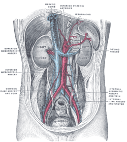 | |
| Posterior abdominal wall, after removal of the peritoneum, showing kidneys, suprarenal capsules, and great vessels. (Hepatic veins labeled at center top.) | |
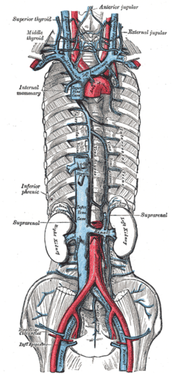 | |
| Superior vena cava, inferior vena cava (IVC), azygos vein and their tributaries. The hepatic veins are seen on the superior portion of the IVC, shortly before it flows into the right atrium, which is not shown. | |
| Latin | venae hepaticae |
| Gray's | p.680 |
| Drains to | inferior vena cava |
| Artery | Hepatic artery |
| Precursor | vitelline veins |
- This page refers to the veins that drain deoxygenated blood from the liver. For the vein that drains nutrient-rich blood from the GI tract and the spleen into the liver, see hepatic portal vein.
In human anatomy, the hepatic veins are the blood vessels that drain de-oxygenated blood from the liver and blood cleaned by the liver (from the stomach, pancreas, small intestine and colon) into the inferior vena cava.
They arise from the substance of the liver, more specifically the central vein of the liver lobule. None of the hepatic veins have valves.
Groups
They can be differentiated into two groups, the upper group and lower group.
- The upper group typically arises from the posterior aspect of the liver, are three in number, and drain the quadrate lobe and left lobe.[citation needed]
- The lower group arise from the right lobe and caudate lobe, are variable in number, and are typically smaller than those in the upper group.[citation needed]
Pathology
Occlusion of the hepatic veins is known as Budd-Chiari syndrome.
External links
- Hepatic Histology: The Lobule - Describes the liver lobule and central vein.
- Hepatic veins - definition - medterms.com
- Hepatic+veins at eMedicine Dictionary
Images of the hepatic veins
- Hepatic veins - Ultrasound - University of the Health Sciences in Bethesda, Maryland
Additional images
-
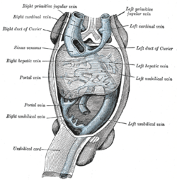
Human embryo with heart and anterior body-wall removed to show the sinus venosus and its tributaries.
-

The portal vein and its tributaries.
-

Longitudinal section of a hepatic vein.
-

Hepatic vein
| ||||||||||||||||||||||||||||||||
| |||||||||||||||||||||||||||||||||||||||||||||||||