Goblet cell
| Goblet cell | |
|---|---|
 | |
| Schematic illustration of a goblet cell in close up, illustrating different internal structures of the cell. | |
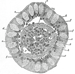 | |
| Transverse section of a villus, from the human intestine. X 350. a. Basement membrane, here somewhat shrunken away from the epithelium. b. Lacteal. c. Columnar epithelium. d. Its striated border. e. Goblet cells. f. Leucocytes in epithelium. f’. Leucocytes below epithelium. g. Blood vessels. h. Muscle cells cut across. | |
| Latin | exocrimohsinocytus caliciformis |
| Code | TH H3.04.03.0.00009; H3.04.03.0.00016 H3.05.00.0.00006 |
Goblet cells are glandular simple columnar epithelial cells whose function is to secrete mucin, which dissolves in water to form mucus. They use both apocrine and merocrine methods for secretion.
The majority of the cell's cytoplasm is occupied by mucinogen granules, except at the bottom. Rough endoplasmic reticulum, mitochondria, the nucleus, and other organelles are concentrated in the basal portion. The apical plasma membrane projects microvilli to increase surface area for secretion.
Locations
They are found scattered among the epithelial lining of organs, such as the intestinal and respiratory tracts.[1] They are found inside the trachea, bronchus, and larger bronchioles in respiratory tract, small intestines, the colon, and conjunctiva in the upper eyelid.(Goblet cells are the chief source of tear mucus. These occur throughout the conjunctiva, especially the plica semilunaris. These are most dense in nasal conjunctiva, least dense in upper temporal fornix and absent in palpebral mucocutaneous junction and limbus.)
They may be an indication of metaplasia, such as in Barrett's esophagus.
Histology
In mucicarmine stains, deep red mucin found within goblet cell bodies.
The nuclei of goblet cells tend to be displaced toward the basal end of the cell body, leading to intense basophilic staining.
Etymology
The term goblet refers to these cells' goblet-like shape. The apical portion is shaped like a cup, as it is distended by abundant mucinogen granules; its basal portion is shaped like a stem, as it is narrow for lack of these granules.
There are other cells that secrete mucus (as in the foveolar cells of the stomach[2]), but they are not usually called "goblet cells" because they do not have this distinctive shape.
Basal secretion
This is the normal base level secretion of mucus, which is accomplished by cytoskeletal movement of secretory granules.
Stimulated secretion
Secretion may be stimulated by dust, smoke, etc.
Other stimuli include viruses, bacteria, etc.
Role in Oral Tolerance
Oral tolerance is the process by which the immune system is prevented from responding to antigen derived from food products, as peptides from food may pass into the bloodstream via the gut, which would in theory lead to an immune response. A recent paper published in Nature, has shed some light on the process and implicated goblet cells as having a role in the process.[3] It was known that CD103 expressing dendritic cells of the lamina propria had a role to play in the induction of oral tolerance (potentially by inducing the differentiation of regulatory T cells), and this paper suggests that the goblet cells act to preferentially deliver antigen to these CD103+ dendritic cells.[3]
See also
- Goblet cell carcinoid - a tumor that has a component that is similar to goblet cells
References
- ↑ "goblet cell" at Dorland's Medical Dictionary
- ↑ BU Histology Learning System: 11303loa - Digestive System: Alimentary Canal: fundic stomach, gastric glands, lumen"
- ↑ 3.0 3.1 McDole et al. (2012). "Goblet cells deliver luminal antigen to CD103+ dendritic cells in the small intestine". Nature 483: 345–349. doi:10.1038/nature10863. PMID 22422267.
Additional images
-

An intestinal gland from the human intestine.
-

Goblet cell in ileum
-

section of mouse intestine. Mucus of goblet cells in blue.
-

Section of mucus membrane of human stomach, near the cardiac orifice. X 45.
c. Cardiac glands.
d. Their ducts.
cr. Gland similar to the intestinal glands, with goblet cells.
mm. Mucous membrane.
m. Muscularis mucosae.
m’. Muscular tissue within the mucous membrane.
External links
- Histology at KUMC epithel-epith08 "Slide 8: Trachea"
- Goblet+cell at eMedicine Dictionary
- Goblet Cells at cvmbs.colostate.edu
- Diagram at uwlax.edu
- Bioweb at UWLAX Zoolab
| ||||||||||||||||||||||||||||||||||||||||||||||||||||||||||
| |||||||||||||||||||||||||||||||||||||||||||||||||||
| ||||||||||||||||||||||||||||||