Ganglioside
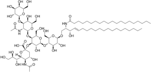
A ganglioside is a molecule composed of a glycosphingolipid (ceramide and oligosaccharide) with one or more sialic acids (e.g. n-acetylneuraminic acid, NANA) linked on the sugar chain. NeuNAc, an acetylated derivative of the carbohydrate sialic acid, makes the head groups of gangliosides anionic at pH 7, which distinguishes them from globosides.
The name ganglioside was first applied by the German scientist Ernst Klenk in 1942 to lipids newly isolated from ganglion cells of the brain.[1] More than 60 gangliosides are known, which differ from each other mainly in the position and number of NANA residues. It is a component of the cell plasma membrane that modulates cell signal transduction events, and appears to concentrate in lipid rafts.
Recently, gangliosides have been found to be highly important molecules in immunology. Natural and semisynthetic gangliosides are considered possible therapeutics for neurodegenerative disorders.[2]
Location
Gangliosides are present and concentrated on cell surfaces, with the two hydrocarbon chains of the ceramide moiety embedded in the plasma membrane and the oligosaccharides located on the extracellular surface, where they present points of recognition for extracellular molecules or surfaces of neighboring cells. They are found predominantly in the nervous system where they constitute 6% of all phospholipids.[3]
Function
The oligosaccharide groups on gangliosides extend well beyond the surfaces of the cell membranes, and act as distinguishing surface markers that can serve as specific determinants in cellular recognition and cell-to-cell communication. These carbohydrate head groups also act as specific receptors for certain pituitary glycoprotein hormones and certain bacterial protein toxins such as cholera toxin.
The functions of gangliosides as specific determinants suggest its important role in the growth and differentiation of tissues as well as in carcinogenesis. It has been found that tumor formation can induce the synthesis of a new complement of ganglioside, and very low concentrations of a specific ganglioside can induce differentiation of cultured neuronal tumor cells.[4]
Common gangliosides

- Two NANAs ("D")
- GD1a
- GD1b
- GD2
- GD3
- Three NANAs ("T")
- GT1b
- Four NANAs ("Q")
- GQ1
Structures of the common gangliosides
GM2-1 = aNeu5Ac(2-3)bDGalp(1-?)bDGalNAc(1-?)bDGalNAc(1-?)bDGlcp(1-1)Cer
GM3 = aNeu5Ac(2-3)bDGalp(1-4)bDGlcp(1-1)Cer
GM2,GM2a(?) = bDGalpNAc(1-4)[aNeu5Ac(2-3)]bDGalp(1-4)bDGlcp(1-1)Cer
GM2b(?) = aNeu5Ac(2-8)aNeu5Ac(2-3)bDGalp(1-4)bDGlcp(1-1)Cer
GM1,GM1a = bDGalp(1-3)bDGalNAc[aNeu5Ac(2-3)]bDGalp(1-4)bDGlcp(1-1)Cer
asialo-GM1,GA1 = bDGalp(1-3)bDGalpNAc(1-4)bDGalp(1-4)bDGlcp(1-1)Cer
asialo-GM2,GA2 = bDGalpNAc(1-4)bDGalp(1-4)bDGlcp(1-1)Cer
GM1b = aNeu5Ac(2-3)bDGalp(1-3)bDGalNAc(1-4)bDGalp(1-4)bDGlcp(1-1)Cer
GD3 = aNeu5Ac(2-8)aNeu5Ac(2-3)bDGalp(1-4)bDGlcp(1-1)Cer
GD2 = bDGalpNAc(1-4)[aNeu5Ac(2-8)aNeu5Ac(2-3)]bDGalp(1-4)bDGlcp(1-1)Cer
GD1a = aNeu5Ac(2-3)bDGalp(1-3)bDGalNAc(1-4)[aNeu5Ac(2-3)]bDGalp(1-4)bDGlcp(1-1)Cer
GD1alpha = aNeu5Ac(2-3)bDGalp(1-3)bDGalNAc(1-4)[aNeu5Ac(2-6)]bDGalp(1-4)bDGlcp(1-1)Cer
GD1b = bDGalp(1-3)bDGalNAc(1-4)[aNeu5Ac(2-8)aNeu5Ac(2-3)]bDGalp(1-4)bDGlcp(1-1)Cer
GT1a = aNeu5Ac(2-8)aNeu5Ac(2-3)bDGalp(1-3)bDGalNAc(1-4)[aNeu5Ac(2-3)]bDGalp(1-4)bDGlcp(1-1)Cer
GT1,GT1b = aNeu5Ac(2-3)bDGalp(1-3)bDGalNAc(1-4)[aNeu5Ac(2-8)aNeu5Ac(2-3)]bDGalp(1-4)bDGlcp(1-1)Cer
OAc-GT1b = aNeu5Ac(2-3)bDGalp(1-3)bDGalNAc(1-4)aXNeu5Ac9Ac(2-8)aNeu5Ac(2-3)]bDGalp(1-4)bDGlcp(1-1)Cer
GT1c = bDGalp(1-3)bDGalNAc(1-4)[aNeu5Ac(2-8)aNeu5Ac(2-8)aNeu5Ac(2-3)]bDGalp(1-4)bDGlcp(1-1)Cer
GT3 = aNeu5Ac(2-8)aNeu5Ac(2-8)aNeu5Ac(2-3)bDGal(1-4)bDGlc(1-1)Cer
GQ1b = aNeu5Ac(2-8)aNeu5Ac(2-3)bDGalp(1-3)bDGalNAc(1-4)[aNeu5Ac(2-8)aNeu5Ac(2-3)]bDGalp(1-4)bDGlcp(1-1)Cer
GGal = aNeu5Ac(2-3)bDGalp(1-1)Cer
where
aNeu5Ac = 5-acetyl-alpha-neuraminic acid
aNeu5Ac9Ac = 5,9-diacetyl-alpha-neuraminic acid
bDGalp = beta-D-galactopyranose
bDGalpNAc = N-acetyl-beta-D-galactopyranose
bDGlcp = beta-D-glucopyranose
Cer = ceramide (general N-acylated sphingoid)
Pathology
Gangliosides are continuously synthesized and degraded in cells. They are degraded to ceramides by sequential removal of sugar units in the oligosaccharide group, catalyzed by a set of highly specific lysosomal enzymes. Mutations in genes coding for these enzymes leads to the accumulation of partially broken down gangliosides in lysosomes, which results in a group of diseases called gangliosidosis. For example, the fatal Tay-Sachs disease arises as genetic defects leads to no functional hexosaminidase A produced, causing GM2 to accumulate in lysosomes, ultimately the ganglion cells in the nervous system swells enormously, disturbing the normal functions of neurons.[3]

Ganglioside are also involved in several diseases:
- Influenza, in which haemagglutinin of influenza virus exploits certain gangliosides to enter and infect the cells expressing them.
- Guillain-Barré syndrome, which has been linked to the production of anti-ganglioside antibodies.[5]
- Cholera
- Tetanus
- Botulism
References
- ↑ "Gangliosides, structure, occurrence, biology and analysis". Lipid Library. The American Oil Chemists' Society.
- ↑ Mocchetti I (2005). "Exogenous gangliosides, neuronal plasticity and repair, and the neurotrophins". Cell Mol Life Sci 62 (19–20): 2283–94. doi:10.1007/s00018-005-5188-y. PMID 16158191.
- ↑ 3.0 3.1 Lubert Stryer (1975). "Biosynthesis of Macromolecular Precursors". Biochemsitry. W H Freeman & Co. p. 486. ISBN 0-7167-0174-X.
- ↑ David L. Nelson; Michael M. Cox (2005). "Lipids". Lehninger Principles of Biochemistry, 4th edition. W H Freeman & Co. p. 357. ISBN 9780716743392.
- ↑ Nachamkin I; Shadomy, SV; Moran, AP; Cox, N; Fitzgerald, C; Ung, H; Corcoran, AT; Iskander, JK et al. (2008). "Anti-ganglioside antibody induction by swine (A/NJ/1976/H1N1) and other influenza vaccines: insights into vaccine-associated Guillain-Barré syndrome". J. Infect Dis. 198 (2): 226–33. doi:10.1086/589624+10.1086/589624. PMID 18522505.
External links
- Gangliosides at the US National Library of Medicine Medical Subject Headings (MeSH)
- Overview of gangliosides at lipidlibrary.co.uk
- Overview of gangliosides at cyberlipid.org
| ||||||||||||||||||||||||||||||
| |||||||||||||||||||||||